- Home
- »
- Healthcare IT
- »
-
AI In Microscopy Market Size & Share, Industry Report, 2033GVR Report cover
![AI In Microscopy Market Size, Share & Trends Report]()
AI In Microscopy Market (2026 - 2033) Size, Share & Trends Analysis Report By Microscopy Type (Optical Microscopy, Fluorescence Microscopy, Electron Microscopy), By Component, By End-use, By Region, And Segment Forecasts
- Report Summary
- Table of Contents
- Segmentation
- Methodology
- Download FREE Sample
-
 Download Sample Report
- Buy Now
Market Size, 2025
$1.1BMarket Estimate, 2026
$1.3BMarket Forecast, 2033
$3.4BCAGR, 2026–2033
14.8%AI In Microscopy Market Summary
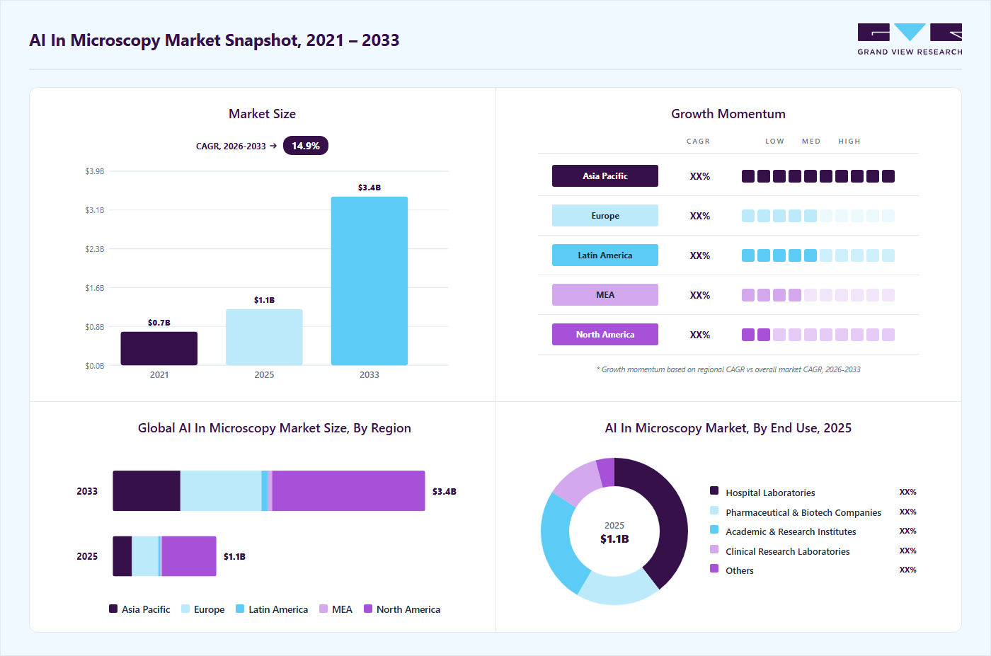
The global AI in microscopy market size was estimated at USD 1.12 billion in 2025 and is projected to reach USD 3.38 billion by 2033, growing at a CAGR of 14.83% from 2026 to 2033. Rising adoption from laboratories and research institutions increasingly seeks faster, more accurate, and reproducible image analysis capabilities.
Key Market Trends & Insights
- North America AI in microscopy market accounted for the largest revenue share of 52.65% in 2025.
- The U.S. AI in microscopy market held the largest market share in 2025.
- Based on microscopy type, optical microscopy segment held the largest market share of 48.41% in 2025.
- Based on component, the AI-enabled microscopes (Hardware) segment accounted for the largest revenue share of 48.19% in 2025.
- Based on end-use, the hospitals segment held the largest market share of 39.47% in 2025.
Market Size & Forecast
- 2025 Market Size: USD 1.12 Billion
- 2033 Projected Market Size: USD 3.38 Billion
- CAGR (2026-2033): 14.83%
- North America: Largest market in 2025
- Asia Pacific: Fastest growing market
The growing complexity and volume of microscopy data, particularly in life sciences, pathology, and materials research, is driving the need for automated and intelligent image interpretation. Advances in deep learning based image recognition, combined with improvements in computing power, are enabling real-time analysis, automated cell classification, and enhanced image resolution. Moreover, rising adoption of high-throughput screening, expanding biomedical research activities, and the need to reduce human error and inter-observer variability are further supporting market growth.")
Growing adoption from laboratories and research institutions drives the growth of the market. A good example of this trend can be seen in the development and deployment of AI‑driven microscopy workflows that enable autonomous data acquisition and analysis, significantly speeding up experimental processes and reducing reliance on manual intervention. For instance, in October 2023, Researchers at the U.S. Department of Energy’s Argonne National Laboratory have pioneered a “self‑driving” microscopy technique in which AI algorithms guide the microscope to focus on regions of interest during scanning, dramatically accelerating data collection and enabling researchers to extract meaningful information without constant human supervision. This kind of autonomous microscopy improves efficiency allows research facilities to handle larger and more complex datasets than traditional methods would permit, illustrating how AI adoption is reshaping laboratory imaging workflows.
Increasing complexity of microscopy data drives adoption of AI‑based analysis fuels growth of the market. According to the ScienceDaily article published in February 2025, microscopy generates increasingly large and complex datasets. Researchers are adopting AI‑based segmentation tools that can automatically identify and outline cells and subcellular structures tasks that would take weeks to perform manually. The international team led by the University of Göttingen retrained an AI model on more than 17,000 annotated microscopy images to create a software called μSAM, which can precisely segment tissues, cells, and even organelles in both light and electron microscopy without extensive manual input. This tool has already been applied in projects ranging from nerve cell analysis in hearing research to automatic tumor cell segmentation for cancer studies, illustrating how AI is helping scientists manage the complexity and volume of modern microscopy data.
"Analysing cells or other structures is one of the most challenging tasks for researchers working in microscopy and is an important task for both basic research in biology and medical diagnostics,""My group specializes in building tools to automate such tasks and we often get asked by researchers to help. Before the development of Segment Anything for Microscopy, we had to ask them to first annotate a lot of structures by hand - a difficult and time-consuming task. μSAM has changed this! Tasks that used to take weeks of painstaking manual effort can be automated in a few hours, because the model can segment any kind of biological structure with a few clicks and can then be further improved to automate the task with our tool. This enables many new applications, and we have already used it in a wide range of projects, ranging from basic cell biology to developing tools for treatment recommendation in cancer therapies."
-Junior Professor Constantin Pape at Göttingen University's Institute of Computer Science
Government initiatives and funding are driving the growth of the Artificial intelligence in microscopy market. For instance, in July 2025, the U.S. National Science Foundation (NSF), awarded a USD 1.2 million grant to researchers at Vanderbilt University to develop an AI-enabled innovative microscope system designed to improve real-time analysis of cellular behaviour in diseases like cancer, automating complex image processing tasks that are difficult or slow to perform manually. The Intelligent Cyber Microscopy System (iCMS), funded by this grant, utilizes adaptive learning and automated decision-making during image acquisition to highlight biologically essential features as they occur, thereby significantly reducing the need for post-processing and accelerating discovery-driven research. This type of government-backed investment enables laboratories and institutions to adopt cutting-edge AI microscopy tools and workflows that can handle large, complex datasets more efficiently, demonstrating how funding agencies are actively supporting innovation in intelligent microscopy technologies.
Recent Funding and Investment for AI In Microscopy
Date
Funding / Investment
Company
News
In October 2025
USD 3.54 (€3 M)
Cytely
Raised to expand its smart AI‑powered microscopy platform that automates cell image analysis and speeds up lab workflows.
In July 2025
USD 10 million
Scopio Labs
Additional funding to scale AI‑based digital microscopy blood cell analysis systems, significantly automating high‑resolution imaging diagnostics.
In May 2025
USD 1.18 million (€1 M)
Genoa Instruments
Funding to advance super‑resolution optical microscopy technologies with next‑gen imaging platforms.
Case Study - AI‑Enhanced High‑Content Microscopy in Infection Research
Case- According to the article published in May 2024, researchers at the London School of Hygiene & Tropical Medicine integrated AI into high‑content microscopy workflows to study Shigella infection in zebrafish models.
Key Points-
-
The team used ZEISS arivis Cloud platform combined with ZEISS Celldiscoverer 7 high‑content system to automate image acquisition and analysis.
-
AI models were trained to detect regions of interest and specific anatomical features across large datasets in 96‑well plate experiments.
-
Automation enabled high-throughput imaging, consistent data acquisition, and reduced manual intervention.
-
The workflow allowed time-series analysis and improved insights into cellular and developmental processes during infection.
Although zebrafish are optically accessible for in vivo imaging of Shigella infection, they are still whole animals. This makes studies involving high-throughput and high-resolution microscopy challenging and time-consuming. With Celldiscoverer 7 and the arivis AI-enhanced workflow, we can image and analyze a lot more samples at a much faster pace.
-Dr. Serge MostowyPrincipal Investigator and Research Team Lead at the Department of Infection Biology, London School of Hygiene & Tropical Medicine, UK
Key Aspect
Market Impact
Accelerated Research
AI-driven automation allows labs to handle larger datasets faster, speeding up experimental timelines.
Improved Reproducibility
Reduces human error and variability, increasing confidence in microscopy-based research findings.
Scalability for Commercial Use
High-throughput, AI-enabled microscopy workflows are attractive for biotech, pharma, and diagnostic markets, driving adoption of AI microscopy platforms.
Market Dynamics
The increasing shift from conventional microscopy to AI-enabled digital pathology platforms is a major driver for the AI in microscopy market. Healthcare providers, research institutions, and pharmaceutical companies are adopting automated imaging systems to improve workflow efficiency, accelerate image interpretation, and reduce manual diagnostic variability. AI-powered microscopy supports automated cell detection, tissue classification, and anomaly identification, enabling faster and more accurate microscopic analysis across pathology, hematology, microbiology, and cytology applications. Growing investments in digital laboratory infrastructure, integrated imaging platforms, and advanced deep learning technologies are accelerating the adoption of AI-assisted microscopy systems across clinical diagnostics and biomedical research environments. For instance, in February 2026, researchers from North Carolina State University developed an AI tool that tracks cells across complete life cycles in live-cell microscopy. It follows cells in 3D over time, from small dishes to complex developing embryos, reducing tracking errors and speeding analysis.
Furthermore, AI-enabled microscopy is also improving diagnostic consistency and supporting large-scale image analysis in laboratories handling increasing testing volumes. Advanced deep learning algorithms and computer vision technologies enhance the detection of tumor cells, blood abnormalities, microbial pathogens, and tissue irregularities with higher analytical precision. Integration of AI with cloud-based imaging platforms and automated workflow systems is strengthening real-time collaboration, remote diagnostics, and data-driven clinical decision-making. Pharmaceutical and biotechnology companies are increasingly utilizing AI-powered microscopy for drug discovery, biomarker identification, and cellular analysis to improve research productivity and reduce development timelines. These technological advancements are expanding AI's role in microscopy across precision medicine, translational research, and next-generation diagnostics.
The high cost of AI-integrated microscopy platforms remains a major barrier to widespread adoption across healthcare institutions, diagnostic laboratories, and research organizations. Implementation of AI-powered microscopy systems requires substantial investment in advanced imaging hardware, high-resolution whole-slide scanners, AI-enabled analytical software, cloud computing infrastructure, and large-scale digital storage systems capable of handling massive microscopy image datasets. Many laboratories transitioning from conventional microscopy to digital AI-assisted workflows must also invest in upgraded networking infrastructure, GPU-enabled servers, cybersecurity systems, and interoperability solutions to support real-time image processing and AI model deployment. These combined infrastructure requirements significantly increase the total cost of ownership for AI microscopy platforms. For instance, according to a comparative developmental and resource-allocation report published in the PubMed Central Journal in January 2023, commercial high-throughput whole slide imaging (WSI) hardware systematically scales from USD 30,000 to over USD 250,000 per unit.
In addition to initial capital expenditure, ongoing operational costs further restrict adoption. Healthcare providers and research institutions must allocate budgets for software licensing subscriptions, AI algorithm updates, cloud storage expansion, technical maintenance, calibration services, and compliance-related validation processes. Integration of AI microscopy systems with laboratory information management systems (LIMS), electronic health records (EHRs), and digital pathology platforms often requires customized IT development and workflow redesign, increasing implementation complexity and associated costs. Staff training programs for pathologists, laboratory technicians, and IT personnel also increase operational expenditures during digital transformation initiatives.
Market Concentration & Characteristics
The chart below illustrates the relationship between industry concentration, industry characteristics, and industry participants. The x-axis represents the level of industry concentration, ranging from low to high. The y-axis represents various industry characteristics, including industry competition, level of partnerships & collaboration activities, degree of innovation, impact of regulations, and regional expansion. The Artificial intelligence in microscopy market is slightly consolidated. However, several emerging players are entering the market, thereby contributing to increased fragmentation within the market. The degree of innovation is high. The level of merger & acquisition activities is moderate. Moreover, the impact of regulations and the regional expansion of industry is high.
AI in microscopy market is driven by continuous innovation, with companies introducing advanced platforms that automate complex imaging tasks, improve accuracy, and streamline laboratory workflows. Prominent players are launching solutions to maintain competitive advantage, while regulatory support further boosts adoption. For example, in February 2025, Honeywell developed Digital Holographic Microscopy (DHM) integrated with AI to automatically count and classify cells and particles. This technology accelerates analysis, reduces manual workload, and can be applied across multiple industries, highlighting how AI is enhancing efficiency and transforming microscopy practices.

“We are seeing a growing need for solutions that can provide results that are both rapid and precise, empowering healthcare professionals to make informed decisions quickly,” “We have invested in innovations like Digital Holographic Microscopy because of their power to make testing and analysis more accessible to patients while also improving patient care and efficiency across our healthcare system.”
-Sarah Martin, president of Honeywell Sensing Solutions
The AI in microscopy industry is experiencing a moderate yet strategic level of merger and acquisition activity as key players seek to enhance technological capabilities, expand product portfolios, and strengthen their position in a rapidly growing market. These moves are driven by the desire to gain competitive advantage, integrate AI‑enabled imaging solutions, and consolidate expertise in microscopy‑based diagnostics and research tools. For example, in July 2025, ZEISS Research Microscopy Solutions acquired all equity shares of Pi Imaging Technology SA, strengthening its access to advanced single‑photon avalanche diode (SPAD) sensor technology for high‑sensitivity imaging that can be integrated into next‑generation microscopy platforms.
Regulations such as HIPAA in the U.S. and GDPR in Europe ensure that AI-powered microscopy platforms handle sensitive imaging and patient data securely. Compliance protects privacy, prevents unauthorized access, and builds trust, enabling broader adoption of AI-based microscopy in research and clinical workflows.
The Artificial intelligence in microscopy market is experiencing significant geographical expansion as key players extend their presence into new regions to capture emerging demand and support local research and clinical needs. Companies are forging strategic partnerships and launching offerings across multiple continents to broaden adoption of AI‑enabled imaging solutions. For instance, in September 2025, Mindpeak, a global provider of AI‑driven pathology and microscopy image analysis tools, recently launched its advanced multiplex immunofluorescence (mIF) analysis solution, PhenoScout AI, in Europe, debuting the platform at Oxford Global’s Precision Medicine Conference in London.
Microscopy Type Insights
Based on microscopy type, optical microscopy segment held the largest market share of 48.41% in 2025, driven by the need for faster, higher-resolution imaging and AI-powered data analysis, allowing researchers to capture and interpret complex biological processes more efficiently. For instance, in March 2025, ZEISS’s Lightfield 4D Optical Microscopy system, for the LSM 910 and LSM 990 confocal platforms. Using the light-field principle, it allows instant volumetric optical imaging with AI-assisted reconstruction, enhancing spatiotemporal accuracy and reducing manual workload in research areas such as neuroscience, cancer biology, and developmental.
The electron microscopy segment is expected to grow at the fastest CAGR during the forecast period, due to laboratories adopt automated and AI‑enhanced workflows to handle increasingly large and complex nanoscale datasets, reducing manual effort and improving throughput. For instance, in March 2025, ZEISS’s release of the ZEN core software for scanning electron microscopes (SEMs), which integrates SEM imaging, EDS analytics, and AI‑assisted workflow enhancements under a single, intuitive interface. By streamlining image acquisition, analysis, and data management across electron microscopes, ZEN core enables both novice and expert users to perform multi‑modal experiments more efficiently and reliably, marking a shift toward connected and intelligent electron microscopy systems that boost productivity in materials science, electronics, and life‑science research.
Component Insights
Based on component, the AI-enabled microscopes (Hardware) segment accounted for the largest revenue share of 48.19% in 2025. Manufacturers embed intelligent analysis directly into microscope systems, allowing real‑time decision support, automated inspection, and greater workflow efficiency. For instance in October 2025, TAGARNO’s AI‑powered digital microscopy platform, which has recently been showcased and commercialized for electronics inspection and industrial microscopy applications. The system combines 4K microscopy imaging with built‑in AI models that automatically detect defects and anomalies in materials and components, reducing manual inspection time and increasing throughput. Integrating AI analytics on the hardware itself, TAGARNO’s solution demonstrates how smart microscope platforms are extending beyond research laboratories into quality control and manufacturing environments, enhancing precision and broadening the scope of microscopy use cases.
The AI-based microscopy software segment is anticipated to register fastest growth from 2026 to 2033, due to laboratories and research institutions increasingly rely on intelligent image analysis tools to accelerate data interpretation, improve accuracy, and handle large and complex datasets. For instance, in May 2025, Leica Microsystems’ release of Aivia 15 , an updated AI‑driven software platform that simplifies microscopy image processing with deep learning‑powered 2D and 3D cell segmentation, automated workflow recommendations, and intuitive guided sequences. Aivia 15 allows researchers to quickly obtain reliable quantitative results across diverse applications such as neuroscience and spatial biology without requiring coding expertise, demonstrating how modern software solutions are making advanced AI capabilities accessible and productive for routine microscopy analysis.
End-use Insights
Based on end-use, the hospitals segment held the largest market share of 39.47% in 2025. Hospital laboratories are progressively integrating AI‑enabled microscopy systems to support faster and more reliable diagnoses while reducing dependence on specialized manual interpretation. For instance, in August 2023, of this trend is the deployment of an automated AI‑microscopy malaria diagnostic system at University College London Hospitals (UCLH), where researchers evaluated a microscope combined with AI software to analyze blood samples from travelers returning from malaria‑endemic regions. In this clinical study, the AI‑assisted microscope scanned and processed more than 1,200 blood samples, identifying malaria parasites with performance close to that of expert microscopists, demonstrating how intelligent imaging platforms can aid clinical decision‑making and ease workloads in busy laboratory environments.

The pharmaceutical & biotechnology companies segment is anticipated to grow at the fastest CAGR from 2026 to 2033. Pharmaceutical and biotechnology companies are increasingly incorporating AI‑enhanced microscopy and high‑content imaging into their research workflows to accelerate drug discovery, streamline compound screening, and gain deeper biological insights from complex cellular data. For instance, Recursion Pharmaceuticals, a U.S. biotech that uses high‑throughput microscopy combined with AI analysis to generate and process millions of cellular images each week as part of its drug discovery platform. By applying machine learning models to these large microscopic datasets, Recursion can detect subtle phenotypic changes in cells exposed to genetic and chemical perturbations, enabling the identification of promising therapeutic candidates much earlier in the development pipeline. This approach not only shortens discovery timelines but also uncovers novel biological mechanisms that traditional methods might miss, illustrating how AI‑based microscopy is reshaping R&D processes in the pharmaceutical and biotech sectors.
Regional Insights
North America AI in microscopy market accounted for the largest revenue share of 52.65% in 2025, driven by strong clinical research activity, advanced hospital laboratories, and collaborations that bring intelligent imaging tools into routine healthcare workflows. For instance, in October 2025, Deepcell’s AI‑powered REM‑I platform, developed in the U.S., which combines high‑resolution microscopy with deep learning to perform label‑free single‑cell imaging and morphological analysis for applications in oncology research, immunology, and drug discovery. Enabling rapid cell profiling and real‑time AI analysis without traditional staining, this technology is helping researchers and clinicians extract detailed cellular insights more quickly and efficiently, emphasizing the region’s role in advancing AI‑enabled microscopy in healthcare and life sciences.

U.S. AI In Microscopy Market Trends
The U.S. AI in microscopy market held the largest market share in 2025. The U.S. key center for the adoption and innovation of Artificial intelligence in microscopy, especially in clinical and diagnostic environments where intelligent imaging can improve patient care and laboratory workflows. For instance, in August 2025, BlurryScope, a compact, AI‑powered portable microscope created by researchers at UCLA, which enables rapid, cost‑effective cancer scoring by applying a deep neural network to continuously scanned tissue images, making it significantly more accessible than traditional whole‑slide scanners. This innovation illustrates how U.S. research institutions are leveraging artificial intelligence to make microscopy more affordable and practical for real‑world clinical diagnostics.
Europe AI In Microscopy Market Trends
Europe Artificial intelligence in microscopy market is expected to witness significant growth during the forecast period. This is attributed to the increasingly acceptance of AI‑driven microscopy, supported by collaborative research initiatives and the deployment of intelligent diagnostic tools across clinical and research settings. For instance, in September 2025, MultiplexAI project, an EU‑funded initiative that aims to turn conventional microscopes into AI‑powered diagnostic platforms capable of detecting multiple parasitic diseases directly at the point of care, strengthening diagnostic capacity in both Europe and partner regions. This project highlights how European research infrastructure and funding mechanisms are enabling advanced microscopy applications that enhance accessibility, speed, and accuracy of disease diagnosis using AI.
The UK AI in microscopy market is expected to grow over the forecast period. The adoption of AI‑assisted microscopy and digital pathology solutions is growing in the UK as clinical laboratories aim to improve diagnostic speed and accuracy. For instance, in May 2025, UK‑based Histofy and Source BioScience announced, which is focused on integrating Histofy’s AI‑powered pathology tools into Source BioScience’s extensive laboratory network. This collaboration is designed to accelerate the rollout of AI‑enhanced microscopy and diagnostic workflows, improve efficiency in cancer detection and other pathology services, and support implementation of intelligent image analysis across NHS and private healthcare labs in the UK.
Germany AI in microscopy market is growing over the forecast period. Germany is becoming a notable hub for AI‑driven microscopy research and applications, supported by strong scientific institutions and collaborations that integrate artificial intelligence into advanced imaging workflows. For instance, in October 2025, international research team including scientists from Friedrich‑Schiller‑Universität Jena, which showcased the first fully autonomous microscopy experiment using an AI agent (AILA) capable of planning, executing, and analysing an atomic force microscopy experiment without human intervention. This achievement highlights how German research environments are pushing the boundaries of intelligent microscopy automation, pointing to future possibilities where AI can independently manage complex imaging experiments and improve laboratory throughput and reproducibility.
Asia Pacific AI In Microscopy Market Trends
Asia Pacific Artificial intelligence in microscopy market is expected to grow at the fastest CAGR in the forecast years. The region is rapidly adopting AI‑powered microscopy and digital pathology technologies as healthcare systems, research labs, and diagnostic centres work to improve disease detection and workflow efficiency. For instance, in August 2025, the launch of the Aperio GT 180 digital pathology scanner by Leica Biosystems at the Asia Pacific Imaging Summit, which brings advanced slide scanning with AI‑enhanced features tailored for high‑throughput clinical and research applications across Asia Pacific healthcare institutions. This launch reflects growing regional demand for automated imaging solutions that support faster, more accurate microscopic analysis in cancer diagnostics and other pathology workflows.
India AI in microscopy marketis expanding rapidly, driven by the growing integration of AI into diagnostic and research workflows and technological advanced product launch. For instance, in July 2024, in Medprime Technologies launched the AI‑integrated digital microscopy platform Micalys, designed to combine whole‑slide imaging, remote robotic control, and AI‑assisted diagnosis for histological and cytological analysis, helping laboratories enhance precision and accessibility of pathology services across clinical and research settings.
China is rapidly advancing AI‑led microscopy and digital pathology tools through research innovation and large imaging datasets that enhance diagnostic intelligence. For instance, in July 2024, the creation of PathOrchestra, a versatile AI model capable of analysing whole‑slide digital pathology images from more than 20 organs, performing tasks such as lesion identification, subtype differentiation, and biomarker assessment with high accuracy. This breakthrough reflects China’s move toward AI‑driven microscopy analysis frameworks that significantly reduce manual interpretation time and improve clinical diagnostic processing across cancer and other pathologies.
Latin America AI In Microscopy Market Trends
Latin America Artificial intelligence in microscopy market is anticipated to grow at a significant CAGR over the forecast period. This is attributed to the growing awareness about AI technologies, increasing government spending, and growing advancements in healthcare infrastructure.
In Argentina, advancements in microscopy‑related AI research are emerging alongside broader digital imaging developments. According to the Springer Nature Limited in October 2023, researchers in the country have contributed to the creation of local clinical image repositories including a clinical and dermoscopy image dataset of skin lesions that are being used to train and evaluate AI tools for dermatological and pathology image analysis. Such datasets are valuable for developing and validating machine learning models tailored to Latin American populations, supporting more accurate diagnostics from microscopic images in clinical practice and research.
Middle East and Africa AI In Microscopy Market Trends
Middle East and Africa AI in microscopy market is expected to grow at a significant CAGR over the forecast period. The market is characterized by a dynamic landscape driven by the growing adoption of technologically advanced medical devices, increasing healthcare expenditures and supportive government policies. Significant integration of AI in healthcare technology across the region contributes to market growth further. Countries such as Saudi Arabia and the UAE are integrating AI into healthcare infrastructure under strategic visions such as Saudi Vision 2033 and the UAE National Strategy for Artificial Intelligence 2031.
Key AI In Microscopy Company Insights
Key players operating in the AI in microscopy market are undertaking various initiatives to strengthen their market presence and increase the reach of their products and services. Strategies such as new product launches and partnerships play a key role in propelling market growth.
Key AI In Microscopy Companies:
The following are the leading companies in the AI in microscopy market. These companies collectively hold the largest market share and dictate industry trends.
- Molecular Devices, LLC.
- SigTuple Technologies Pvt. Ltd.
- Leica Microsystems
- Nikon Corporation Healthcare Business Unit
- Revvity Signals Software, Inc.
- Oxford Instruments
- ZEISS
- Thermo Fisher Scientific Inc.
- KOLAIDO GmbH.
- Ariadne.ai ag
Competitive Benchmarking
Operating Strategies
Competitive Edge
Weaknesses
Mature Players: Danaher Corporation
- Mature AI in microscopy companies are focusing on expanding integrated digital pathology and imaging ecosystems that combine AI software, cloud-based analytics, whole-slide imaging, and laboratory workflow automation platforms.
- These players emphasize strategic partnerships with hospitals, pharmaceutical companies, and research institutes.
- Established companies maintain a strong competitive advantage through extensive imaging expertise, large installed microscopy bases, proprietary imaging datasets, and integration capabilities with laboratory information systems and digital pathology platforms.
- Strong global distribution networks, regulatory experience, and long-term collaborations with healthcare institutions support broad commercial adoption and continuous AI.
- High implementation and operational costs associated with AI-enabled microscopy systems, digital scanners, cloud infrastructure, and enterprise software integration may limit adoption among smaller laboratories and healthcare facilities.
- Dependence on high-quality annotated datasets, interoperability challenges with legacy laboratory infrastructure, and lengthy clinical validation processes increase deployment complexity and commercialization timelines.
Emerging Players: Quotient, Ltd.
- Emerging AI microscopy companies are adopting niche-focused strategies targeting AI-assisted cancer detection, automated cellular analysis, biomarker quantification, and pathology workflow optimization to gain market traction.
- Many startups are leveraging cloud-native AI platforms, scalable subscription models, and collaborative research partnerships to improve accessibility and accelerate deployment across clinical and research laboratories.
- Emerging players benefit from agile AI development cycles, advanced deep learning capabilities, and specialization in targeted diagnostic applications that enable rapid innovation and customized solution development.
- Their ability to integrate modern AI architectures and adapt quickly to evolving clinical requirements supports faster deployment of specialized microscopy analytics solutions.
- Emerging players typically face limited brand recognition, smaller distribution networks, and lower financial resources compared with established companies. High regulatory compliance costs, reimbursement challenges, and dependence on partnerships for commercialization may restrict expansion. Limited installed customer bases can also affect recurring revenue generation.
Recent Developments
-
In August 2025, ZEISS and Alpenglow Biosciences announced a collaboration to co‑develop a 3D pathology solution combining inverted light‑sheet microscopy with AI‑driven data processing to enhance clinical and research imaging.
"Partnering with ZEISS represents a significant step forward in our mission to revolutionize pathology by extracting and analysing the wealth of information contained in every single cell of a biopsy, rather than relying on just a small fraction of the tissue,"
-Dr. Nicholas Reder, CEO of Alpenglow Biosciences.
-
In February 2025, Motic Instruments and Media Cybernetics announced a strategic partnership to integrate advanced microscopy hardware with AI‑driven Image‑Pro software for research and clinical imaging.
-
In April 2024, ZEISS partnered with Argolight to integrate microscopy quality control solutions with ZEISS platforms, enhancing reliability and reproducibility of microscopy imaging systems.
AI In Microscopy Market Report Scope
Report Attribute
Details
Market size value in 2026
USD 1.28 billion
Revenue forecast in 2033
USD 3.38 billion
Growth rate
CAGR of 14.83% from 2026 to 2033
Actual data
2021 - 2025
Forecast period
2026 - 2033
Quantitative units
Revenue in USD million/billion and CAGR from 2026 to 2033
Report coverage
Revenue forecast, company ranking, competitive landscape, growth factors, and trends
Segments covered
Microscopy type, component, end-use, region
Regional scope
North America; Europe; Asia Pacific; Latin America; MEA
Country scope
U.S.; Canada; Mexico; Germany; UK; France; Italy; Spain; Denmark; Sweden; Norway; China; Japan; India; South Korea; Australia; Thailand; Brazil; Argentina; South Africa; Saudi Arabia; UAE; Kuwait
Key companies profiled
Molecular Devices, LLC; SigTuple Technologies Pvt. Ltd.; Leica Microsystems; Nikon Corporation Healthcare Business Unit; Revvity Signals Software, Inc.; Oxford Instruments; ZEISS; Thermo Fisher Scientific Inc.; KOLAIDO GmbH.; Ariadne.ai ag
Customization scope
Free report customization (equivalent up to 8 analysts working days) with purchase. Addition or alteration to country, regional & segment scope.
Pricing and purchase options
Avail customized purchase options to meet your exact research needs. Explore purchase options
Global AI In Microscopy Market Report Segmentation
This report forecasts, revenue growth at global, regional, and country levels and provides an analysis of the latest industry trends in each of the sub-segments from 2021 to 2033. For this study, Grand View Research has segmented global AI in microscopy market report based on component, technology, modality, application, end-use, and region.

-
Microscopy Type Outlook (Revenue, USD Million, 2021 - 2033)
-
Optical Microscopy
-
Fluorescence Microscopy
-
Electron Microscopy
-
Others
-
-
Component Outlook (Revenue, USD Million, 2021 - 2033)
-
AI-Enabled Microscopes (Hardware)
-
AI-Based Microscopy Software
-
Services
-
-
End-use Outlook (Revenue, USD Million, 2021 - 2033)
-
Hospital Laboratories
-
Pharmaceutical & Biotechnology Companies
-
Academic & Research Institutes
-
Clinical Research Laboratories
-
Others
-
-
Regional Outlook (Revenue, USD Million, 2021 - 2033)
-
North America
-
U.S.
-
Canada
-
Mexico
-
-
Europe
-
Germany
-
UK
-
France
-
Italy
-
Spain
-
Denmark
-
Sweden
-
Norway
-
-
Asia Pacific
-
China
-
Japan
-
India
-
South Korea
-
Australia
-
Thailand
-
-
Latin America
-
Brazil
-
Argentina
-
-
MEA
-
South Africa
-
Saudi Arabia
-
UAE
-
Kuwait
-
-
Delivered Customizations
This report has been delivered with the following In-depth customizations
Client Request
Customization Delivered
Value Adds
AI-Enabled Digital Pathology & Automated Imaging Analysis
Developed a tailored analysis of the AI in microscopy market focused on adoption of digital pathology platforms, AI-assisted image analysis, automated cellular detection, and integration of deep learning algorithms across pathology, hematology, microbiology, and life sciences research applications. The study incorporated assessment of healthcare digitization trends, laboratory automation adoption, AI-based diagnostic workflows, regulatory developments, and deployment of cloud-enabled microscopy platforms across healthcare and research institutions.
Enables stakeholders to understand evolving AI-driven microscopy ecosystems, identify high-growth diagnostic and research applications, assess infrastructure readiness and laboratory digitization trends, and evaluate the impact of AI-assisted diagnostics on clinical efficiency, research productivity, and long-term market expansion.
Laboratory Automation & AI Microscopy Adoption Trends
Delivered a customized evaluation of laboratory adoption and utilization patterns for AI-enabled microscopy solutions, including trend analysis of digital pathology implementation, automated imaging workflows, cloud-based microscopy platforms, and AI-assisted diagnostic decision support systems. The study also assessed factors influencing AI microscopy adoption across hospitals, diagnostic laboratories, pharmaceutical companies, and academic research institutions.
Provides actionable insights into evolving laboratory automation trends, emerging diagnostic imaging preferences, and commercially attractive application segments, helping clients identify high-demand opportunities and align strategic initiatives with changing healthcare and research requirements.
Healthcare Infrastructure & Precision Diagnostics Opportunity Assessment
Conducted a focused assessment of healthcare provider and research institution demand for AI-integrated microscopy technologies, including pathology workflow modernization, precision diagnostics adoption, computational imaging infrastructure, and AI-supported biomarker analysis capabilities. The analysis also evaluated gaps in digital laboratory infrastructure, interoperability readiness, skilled workforce availability, and AI implementation challenges across developed and emerging healthcare markets.
Supports investment planning for AI microscopy platforms, digital pathology infrastructure, and laboratory automation technologies by quantifying market demand, identifying underserved clinical and research segments, evaluating commercialization potential, and strengthening business expansion strategies across precision medicine and diagnostic imaging ecosystems.
Frequently Asked Questions About This Report
The global AI in microscopy market size was estimated at USD 1.12 billion in 2025 and is expected to reach USD 1.29 billion in 2026.
The global AI in microscopy market is expected to grow at a compound annual growth rate of 14.83% from 2026 to 2033 to reach USD 3.38 billion by 2033.
The optical microscopy segment held the largest market share of 48.41% in 2025.
Some key players operating in the AI in microscopy market include Molecular Devices, LLC ; SigTuple Technologies Pvt. Ltd.; Leica Microsystems; Nikon Corporation Healthcare Business Unit; Revvity Signals Software, Inc.; Oxford Instruments; ZEISS; Thermo Fisher Scientific Inc.; KOLAIDO GmbH.; Ariadne.ai ag
Key factors that are driving the AI in microscopy market are rising adoption of high-throughput screening, expanding biomedical research activities, and the need to reduce human error and inter-observer variability. Advances in deep learning based image recognition, combined with improvements in computing power, are enabling real-time analysis, automated cell classification, and enhanced image resolution.
About the Author(s)
Healthcare IT Research Team
Healthcare · Healthcare ITThis report was authored by the healthcare it research team at Grand View Research - comprising two research analysts, one senior research analyst, and one industry expert - with specialized expertise in the healthcare it segment of the healthcare industry. All findings are based on proprietary healthcare databases, executive interviews, and regulatory analysis, subject to internal peer review prior to publication.
Last Updated:
Speak to Analyst


Share this report with your colleague or friend.
Need a Tailored Report?
Customize this report to your needs — add regions, segments, or data points, with 20% free customization.

ISO 9001:2015 & 27001:2022 Certified
We are GDPR and CCPA compliant! Your transaction & personal information is safe and secure. For more details, please read our privacy policy.