
- Home
- »
- Medical Devices
- »
-
Light-Sheet Fluorescence Microscopy Market Report, 2035GVR Report cover

Light-Sheet Fluorescence Microscopy Market (2027 - 2035)
Size, Share & Trends Analysis Report By Product Type (Light-Sheet Microscopy Systems, Light-Sheet Modules & Retrofit Kits, Accessories & Components), By Application, By Region, And Segment Forecasts
Market Size, 2026
$146.7MMarket Estimate, 2027
$159.0MMarket Forecast, 2035
$317.7MCAGR, 2027–2035
9.0%Light-Sheet Fluorescence Microscopy Market Summary
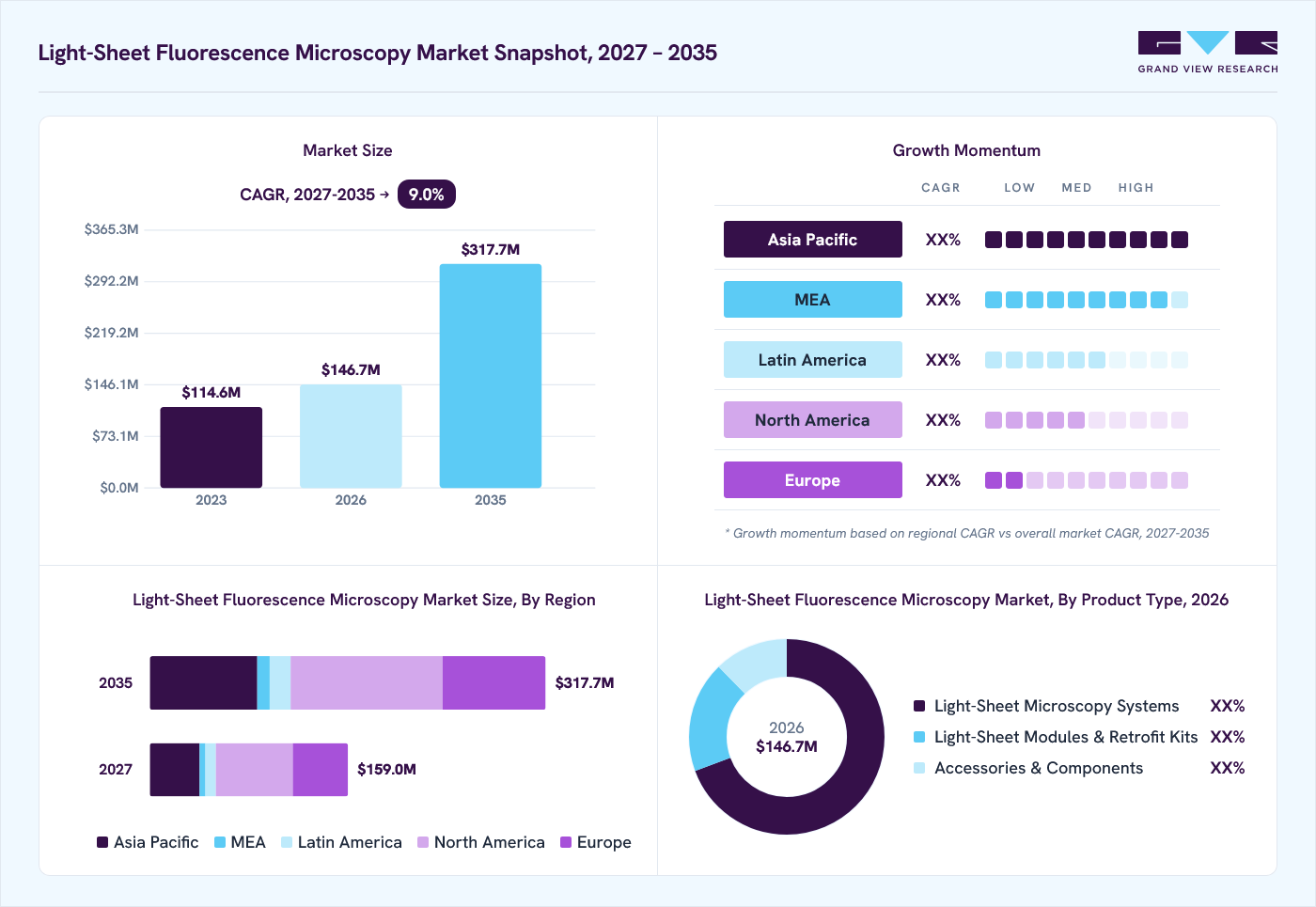
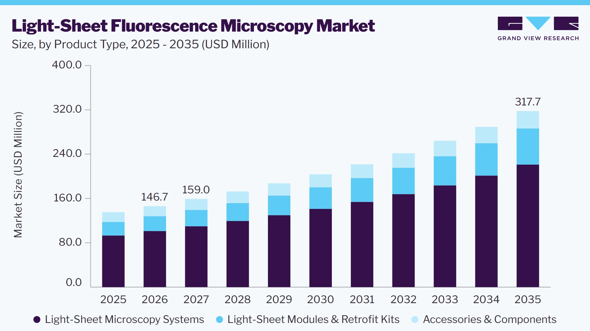
The light-sheet fluorescence microscopy market size was valued at USD 146.7 million in 2026 and is projected to grow from USD 159.0 million in 2027 to USD 317.7 billion by 2035, at a CAGR of 9.0% from 2027 to 2035. North America market dominated market in 2026 and accounted for the largest revenue share of 38.9%. This growth is driven by increasing demand for high-resolution 3D imaging in developmental biology, neuroscience, cancer research, and live-cell imaging. Its ability to minimize phototoxicity and photobleaching while enabling long-term observation of living specimens is supporting adoption.
 Source: Grand View Research, IR Documents, Primary Interviews, Paid DatabasesTo learn more about this report,
 Download Free Sample Report
Key Market Trends & Insights
- By product type: Light-sheet microscopy systems segment held the largest revenue share of 69.3% in 2026.
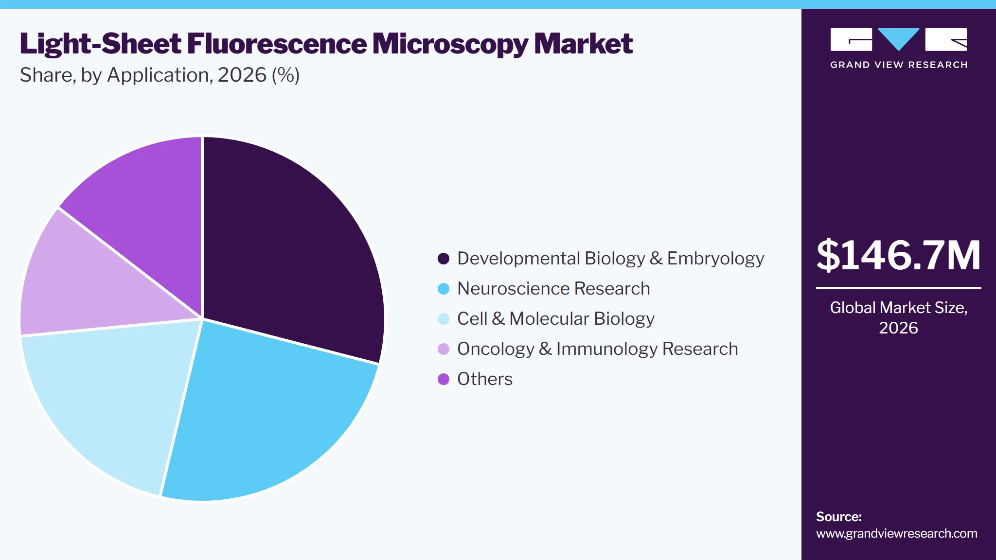
- By application: Developmental biology & embryology segment held the largest revenue share of 29.0% in 2026.
Regional Highlights
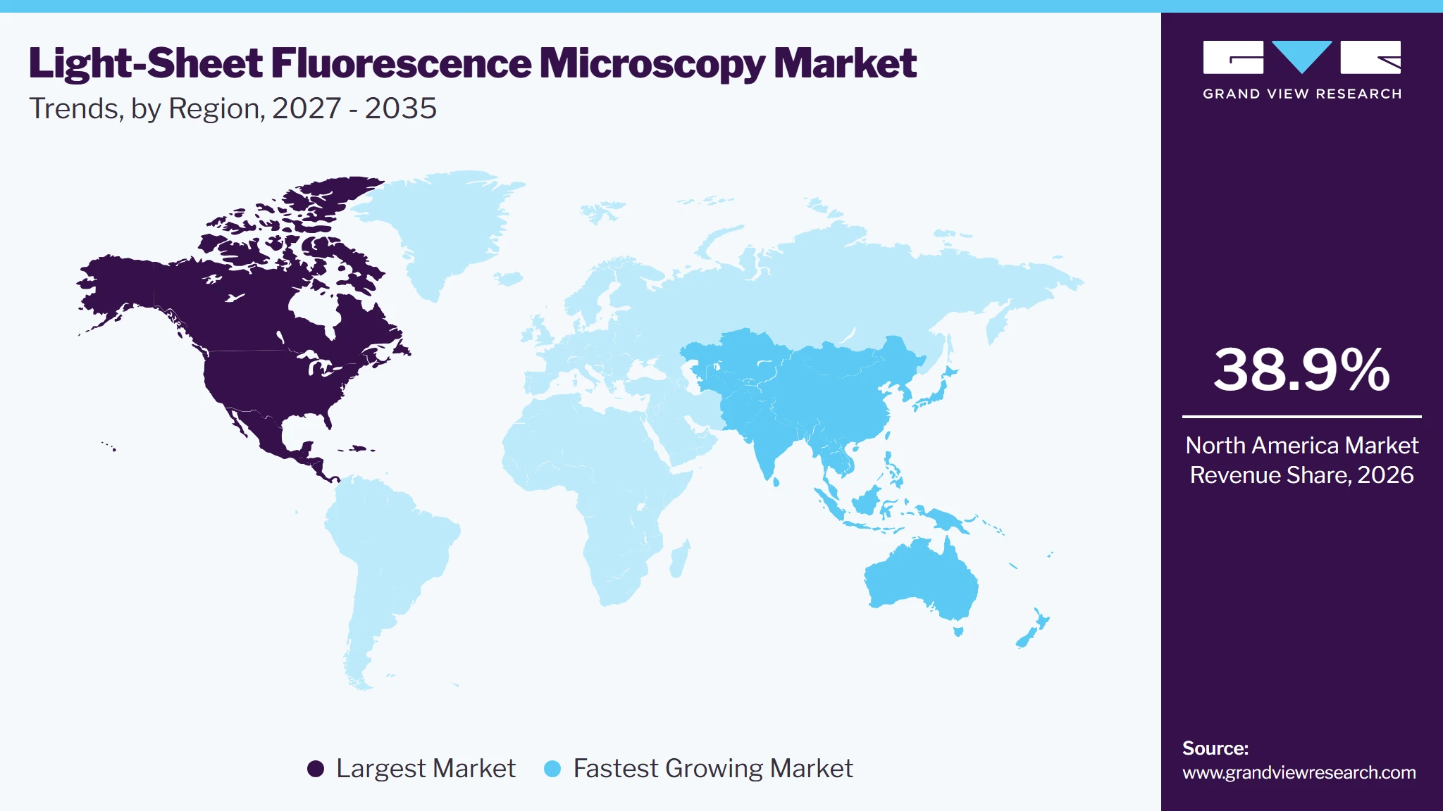
- Largest regional market: North America (38.9% market share, 2026)
- Fastest-growing regional market: Asia Pacific (highest CAGR, 2027-2035)
Market Size & Forecast
- Market size in 2026: USD 146.7 Million
- Estimated market size in 2027: USD 159.0 Million
- Projected market size in 2035: USD 317.7 Million
- CAGR (2027-2035): 9.0%
Prism AI by Grand View Research Free to tryAsk a question about the Light-Sheet fluorescence microscopy marketTry asking✓Every figure cited from GVR research Prefer the report? Download free sample →Growing investments in life sciences research, increasing use of organoids and 3D cell culture models, and technological advancements in automated imaging, AI-based image analysis, and faster image acquisition are further driving market growth.
 To learn more about this report, Download Free Sample Report
Market Dynamics
The industry is witnessing a shift toward high-throughput, low-phototoxicity imaging solutions capable of capturing complex biological structures in three dimensions. Increasing reliance on whole-organ and whole-embryo imaging, organoid research, and long-term live-cell observation is driving demand for systems that offer faster image acquisition with minimal sample damage. Research laboratories and pharmaceutical companies are increasingly adopting advanced light-sheet platforms to investigate disease mechanisms, cellular interactions, developmental processes, and drug responses.
However, high instrument acquisition costs, specialized sample preparation requirements, and the need for skilled operators continue to limit adoption, particularly among smaller research institutions. Market competition is increasingly centered on improving imaging resolution, automation, multi-view imaging, and AI-assisted image reconstruction, while expanding compatibility with diverse biological samples and enabling more reproducible imaging workflows.
The increasing use of organoids and 3D cell culture models in drug discovery, disease modelling, cancer research, and developmental biology is driving demand for light-sheet fluorescence microscopy. Light-sheet microscopy enables researchers to visualize entire organoids, tissues, and complex cellular structures in three dimensions. Its ability to provide rapid volumetric imaging while minimizing phototoxicity and photobleaching is particularly valuable for studying cell differentiation, tissue development, drug responses, and cellular interactions over extended periods. Consequently, growing investments in organoid-based research and advanced preclinical models are expanding the need for sophisticated imaging systems.
Case Study Published by Nature in June 2025: ETH Zurich Advances Long-Term 3D Imaging of Human Brain Organoids Using Light-Sheet Microscopy
Objective: To investigate the cellular and tissue-level dynamics involved in early human brain development using advanced live-cell imaging.
Development: Researchers from ETH Zurich and collaborating institutions established a long-term live light-sheet fluorescence microscopy approach using fluorescently labelled human induced pluripotent stem cell-derived brain organoids. Published in Nature on June 18, 2025, the study enabled researchers to track tissue morphology, cellular behavior, and subcellular structures throughout organoid development. The team conducted imaging over approximately two weeks, including an 188-hour imaging experiment with 30-minute acquisition intervals. The approach also enabled simultaneous monitoring of multiple cellular components, including actin, tubulin, plasma membrane, and nuclear structures.
Market Significance: The study demonstrates the importance of light-sheet fluorescence microscopy in organoid-based research by enabling long-term, high-resolution 3D visualization of living biological samples with minimal phototoxicity. Such applications are expected to support demand for advanced microscopy systems in neuroscience, developmental biology, disease modelling, and pharmaceutical research.
Light-sheet fluorescence microscopy systems require substantial initial investment in specialized lasers, high-sensitivity cameras, precision optics, and dedicated sample preparation equipment. Additional costs associated with system calibration, maintenance, software upgrades, and skilled operators can limit adoption among smaller research laboratories and academic institutions. For instance, commercial systems from manufacturers such as ZEISS and Miltenyi Biotec incorporate specialized illumination and detection components, increasing the investment required compared with conventional fluorescence microscopy.
Increasing use of 3D cellular models, organoids, and tumor spheroids in preclinical drug development is creating opportunities for light-sheet microscopy adoption. The technology enables researchers to visualize drug penetration, cellular responses, and structural changes across intact 3D models, supporting more detailed compound screening and toxicity assessment. For instance, an October 2026 study published in PubMed demonstrated the use of dual-view oblique plane microscopy (dOPM), a light-sheet imaging approach, for high-content screening of small-molecule compounds in 3D glioblastoma stem-cell spheroids, achieving single-cell-level analysis in a 96-well plate format.
Light-sheet fluorescence microscopy requires specialized sample preparation, particularly for large or intact biological specimens, where tissue clearing, labeling, and refractive-index matching are often necessary to obtain high-quality images. These procedures are time-consuming and may affect sample integrity or fluorescence signals. In addition, high-speed 3D imaging generates large volumes of multidimensional image data, requiring substantial storage, computational resources, and advanced image-processing software, which increase operational complexity and limit adoption among smaller research laboratories.
An October 2024 study published in Nature Methods, titled Image processing tools for petabyte-scale light sheet microscopy data, highlighted that individual light-sheet microscopy experiments can generate hundreds of gigabytes of data. The researchers developed PetaKit5D to address limitations in conventional image-processing tools, demonstrating the need for specialized computational infrastructure.
Market Concentration & Characteristics
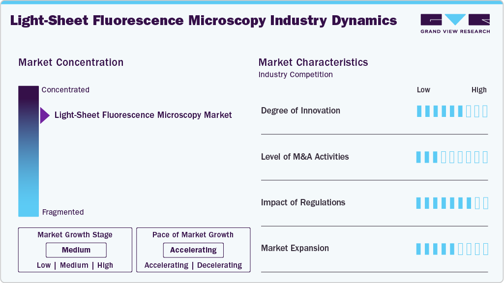
The chart below represents the relationship between industry concentration, industry characteristics, and industry participants. The industry consolidates, with few large manufacturers entering the market. There is a high degree of medium, moderate level of merger & acquisitions activities, high impact of regulations, and moderate high expansion of industry.
The market is witnessing a high degree of innovation, driven by advancements in high-speed volumetric imaging, multi-view imaging, AI-enabled image reconstruction, and improved optical resolution. Manufacturers are developing systems that enable faster imaging of complex biological specimens while minimizing phototoxicity. For instance, in October 2026, researchers introduced the Multi-Immersion Oblique Plane Microscope (miOPM), which supports high-resolution imaging across different sample types using interchangeable oil, water, and air objectives.
 To learn more about this report, Download Free Sample Report
The level of mergers and acquisitions and strategic partnership activity is moderate, as established microscopy manufacturers are acquiring specialized imaging technology developers and collaborating with niche players to strengthen their product portfolios. For instance, in May 2024, Leica Microsystems integrated Viventis Microscopy's light-sheet technology into its portfolio, adding the Viventis LS2 Live system and strengthening its capabilities in volumetric live-cell imaging.
The impact of regulations is moderate, as manufacturers must comply with electrical safety, electromagnetic compatibility (EMC), laser safety, and hazardous-substance restrictions to commercialize microscopy systems across different regions. For instance, Leica Microsystems' Viventis Deep light-sheet microscope received a UK Declaration of Conformity in October 2024, demonstrating compliance with UK electrical safety, EMC, and RoHS requirements.
Market expansion is supported by increasing commercialization, international distribution, and installations across academic research institutions and life sciences laboratories, particularly in North America, Europe, and Asia Pacific. For instance, MBF Bioscience's SLICE light-sheet microscope had been installed at more than 30 research institutions across three continents by May 2026, demonstrating the growing international reach of specialized light-sheet imaging systems.
Analyst Perspective
The light-sheet microscopy market is driven by growing demand for high-resolution, high-speed 3D imaging while minimizing phototoxicity and photobleaching in living biological specimens. Competitive differentiation is increasingly focused on faster volumetric imaging, automated sample handling, improved fluorescence sensitivity, and AI-enabled image analysis. ZEISS, Bruker, Leica Microsystems, Miltenyi Biotec, and Intelligent Imaging Innovations (3i) are positioned in advanced systems, while specialized developers such as PhaseView, Viventis Microscopy, and Applied Scientific Instrumentation (ASI) address application-specific requirements. Significant opportunities exist in automated, high-throughput platforms for organoid research, drug discovery, developmental biology, and large-volume tissue imaging. However, high equipment costs, complex sample preparation, specialized technical requirements, and computational challenges associated with large 3D datasets may continue to limit adoption.
Product Type Insights
The light-sheet microscopy systems segment accounted for the largest market revenue share of 69.3% in 2026 due to the growing preference for fully integrated imaging platforms that offer high-speed 3D imaging, automated sample positioning, and advanced fluorescence detection. These systems are widely adopted by academic institutions, pharmaceutical companies, and life sciences research laboratories for complex biological imaging. For instance, ZEISS Lightsheet 7 provides dedicated light-sheet imaging capabilities for observing living specimens and large cleared tissues, supporting applications in developmental biology and neuroscience.
The light-sheet modules & retrofit kits segment is expected to grow at the fastest CAGR over the forecast period driven by increasing demand for cost-effective upgrades that allow research laboratories to incorporate light-sheet imaging capabilities into existing microscopy infrastructure. Modular solutions reduce the need to purchase entirely new imaging systems and provide flexibility in adapting existing laboratory setups. For instance, openUC2's Light-Sheet Add-On provides a modular approach to implementing light-sheet illumination and 3D imaging capabilities, supporting the adoption of add-on configurations in research laboratories.
Application Insights
The developmental biology & embryology segment accounted for the largest market revenue share of 29.0% in 2026. This is primarily due to the extensive use of light-sheet microscopy to study embryonic development, cell migration, tissue formation, and organogenesis. Its ability to capture dynamic biological processes in living embryos over extended periods with minimal phototoxicity supports its widespread adoption in developmental research. For instance, a July 2024 study published in the Journal of Visualized Experiments (JoVE) demonstrated light-sheet microscopy-based imaging and mounting strategies for observing early zebrafish embryo development over extended periods.
 To learn more about this report, Download Free Sample Report
The oncology & immunology research is expected to grow at the fastest CAGR over the forecast period. This is driven by increasing demand for advanced 3D imaging to investigate tumor microenvironments, immune-cell interactions, cancer progression, and responses to immunotherapy. Light-sheet microscopy enables researchers to visualize immune-cell distribution and tumor architecture across large tissue volumes, supporting cancer research and drug evaluation. For instance, a February 2025 study published in iScience established a combined light-sheet and confocal microscopy workflow to analyze immune responses within mouse primary and metastatic tumors, demonstrating its potential in cancer and immunotherapy research.
Regional Insights
North America dominated the market with a revenue share of 38.9% in 2026 driven by substantial investments in biomedical research, advanced microscopy infrastructure, neuroscience studies, and pharmaceutical R&D. Increasing adoption of high-resolution 3D imaging for studying brain architecture, cellular interactions, and tissue development is further supporting regional demand. In February 2026, Princeton University announced funding for a new LICONN light-sheet fluorescence microscope at its Bezos Center for Neural Circuit Dynamics. The system is intended to support nanoscale imaging of brain architecture, including individual axons and synapses.
 To learn more about this report, Download Free Sample Report
U.S. Light-Sheet Fluorescence Microscopy Market Trends
The U.S. Light-sheet fluorescence microscopy market is driven by increasing NIH funding for advanced imaging infrastructure and growing demand for shared microscopy facilities across academic and biomedical research institutions. In 2025, NIH supported the establishment of a shared light-sheet microscopy facility at Seattle Children's Research Institute using LifeCanvas Technologies' SmartSPIM system. The facility will support research in neurodevelopmental disorders, cancer biology, stem cells, and tissue regeneration.
Europe Light-Sheet Fluorescence Microscopy Market Trends
Europe is witnessing demand for advanced fluorescence imaging technologies, supported by collaborative life sciences research, government-funded scientific infrastructure, and increasing applications in developmental biology, neuroscience, and cellular research. In December 2025, researchers from the University of Göttingen, University Medical Center Göttingen, and the University of Lübeck developed an improved light-sheet fluorescence microscopy platform to enable faster and clearer 3D imaging of large biological samples, including tissues and organs.
The Germany Light-sheet fluorescence microscopy market is supported by investments in university-based imaging facilities and increasing availability of specialized microscopy platforms for live-cell imaging, neuroscience, and molecular biology research. The University of Osnabrück's Integrated Bioimaging Facility provides access to lattice light-sheet microscopy through its Center for Cellular Nanoanalytics, supporting advanced live-cell and fluorescence imaging research.
The UK semiconductor SEM systems market is driven primarily by increasing adoption of advanced imaging systems in cancer research, cell biology, and live-cell analysis. The Institute of Cancer Research, London, operates three light-sheet microscopes within its Chelsea imaging facility. It was also the first UK research organization to acquire a lattice light-sheet microscope, supporting detailed observation of dynamic cellular processes such as cell division.
Asia Pacific Light-Sheet Fluorescence Microscopy Market Trends
Asia Pacific is expected to experience strong growth, supported by expanding biotechnology research, increasing government investments in scientific infrastructure, and growing adoption of advanced microscopy technologies across academic institutions and pharmaceutical research centers. The National University of Singapore operates a dedicated 3i light-sheet microscopy facility at its Centre for BioImaging Sciences, offering live-cell imaging capabilities and isotropic-resolution imaging for biological research.
The China Light-sheet fluorescence microscopy market is supported by growing investments in optical imaging research, biomedical engineering, and the development of advanced microscopy technologies for biological applications. In August 2024, researchers affiliated with Huazhong University of Science and Technology published research highlighting advances in light-sheet fluorescence microscopy, particularly its applications in high-speed 3D imaging, biological discovery, and reduced phototoxicity
The India Light-sheet fluorescence microscopy market is driven by increasing investments in advanced research facilities, growing academic interest in 3D biological imaging, and expanding life sciences research infrastructure. IIT Bombay operates a 3i Lattice Light-Sheet Microscopy Facility that supports 3D and 4D imaging of cells and small organisms, including zebrafish embryos and larvae. The facility is also accessible to external academic and industrial users.
Latin America Light-Sheet Fluorescence Microscopy Market Trends
The Latin America market is primarily driven by growing investments in biomedical research infrastructure, increasing collaborations between universities and international research organizations, and efforts to improve access to advanced bioimaging technologies. The Chan Zuckerberg Initiative supports the Developing a Latin American Light-Sheet Bioimaging Hub project, led by Universidad Mayor in Chile. The initiative connects research laboratories across Chile and international institutions to expand access to light-sheet microscopy through training, workshops, and remote imaging services
The Brazil Light-sheet fluorescence microscopy market is expected to benefit from expanding advanced microscopy infrastructure, increasing investments in biological imaging, and growing demand for specialized research facilities. The Brazile Biosciences National Laboratory (LNBio), part of CNPEM, operates a Biological Imaging Laboratory offering advanced fluorescence and super-resolution microscopy capabilities, supporting research in cellular, molecular, and structural biology.
Middle East & Africa Light-Sheet Fluorescence Microscopy Market Trends
The Middle East & Africa market is supported by increasing investments in academic research infrastructure, expansion of university-based biomedical laboratories, and growing adoption of advanced fluorescence imaging for cellular and molecular biology research. The University of Sharjah's Research Institute for Medical and Health Sciences operates a Molecular Imaging Core Facility equipped with advanced fluorescence microscopy systems. The facility provides imaging capabilities and technical support to researchers and students across UAE universities and research institutions.
The UAE Light-sheet fluorescence microscopy market benefits from expanding biomedical research infrastructure, investments in university research facilities, and increasing demand for advanced imaging technologies in neuroscience, cellular biology, and molecular research. NYU Abu Dhabi's Light Microscopy Core Technology Platform supports fluorescence imaging, 3D neuronal network studies, cell screening, and imaging of biological specimens such as mouse brain sections and cultured cells.
Key Light-sheet Fluorescence Microscopy Company Insights
The Light-sheet fluorescence microscopy market players are focusing on devising innovative business growth strategies in the form of product portfolio expansions, partnerships & collaborations, mergers & acquisitions, and business footprint expansions.
Key Light-sheet Fluorescence Microscopy Companies
The following key companies have been profiled for this study on the light-sheet fluorescence microscopy market.
-
ZEISS
-
Leica Microsystems
-
Bruker
-
Miltenyi Biotec
-
Nikon Corporation Healthcare Business Unit
-
EVIDENT
-
Tecan Trading AG
-
PhaseView
-
Scientifica
Competitive Benchmarking
Category
Operating Strategies
Competitive Edge
Weakness
Established Players (e.g., ZEISS, Leica Microsystems, Bruker, Miltenyi Biotec, Nikon Corporation Healthcare Business Unit, EVIDENT, Tecan Trading AG)
Invest in advanced light-sheet imaging technologies, high-speed fluorescence detection, automated sample handling, multi-view imaging, and AI-enabled image processing. Expand integrated microscopy portfolios through product innovation, application-specific systems, software development, and collaborations with academic and pharmaceutical research institutions to address demand for 3D imaging, organoid research, neuroscience, and drug discovery.
Strong global distribution networks, established relationships with academic and pharmaceutical research organizations, extensive R&D capabilities, recognized microscopy brands, and broad portfolios of optical imaging technologies. Established players can leverage existing customer bases and technical support infrastructure to expand adoption across research applications.
High equipment acquisition and maintenance costs, complex system configurations, and dependence on specialized technical expertise. Competition from alternative imaging technologies, lengthy purchasing cycles at research institutions, and the need for application-specific sample preparation and data-processing capabilities can constrain adoption.
Emerging and Specialized Players (e.g., PhaseView, Scientifica)
Focus on specialized light-sheet configurations, modular imaging platforms, customized illumination systems, and application-specific solutions for neuroscience, developmental biology, and live-cell imaging. Strengthen market presence through collaborations with research laboratories, technology partnerships, customized system development, and improvements in imaging flexibility and workflow efficiency.
Specialized expertise in light-sheet imaging, flexibility in developing application-specific microscopy solutions, and the ability to address niche research requirements. Focused product portfolios enable differentiation through customized imaging configurations, specialized sample compatibility, and research-oriented technical support.
Comparatively narrower product portfolios, limited global distribution and service infrastructure, and lower financial resources than established microscopy manufacturers. Dependence on specialized research applications, customer-specific configurations, and competition from integrated microscopy platforms can restrict broader commercial adoption.
Recent Developments
-
In October 2025, LifeCanvas Technologies launched DALISPIM, an open-top light-sheet microscope designed to streamline high-throughput 3D imaging and spatial biology research. The system incorporates patent-pending Depth Adaptive Light Interface (DALI) technology, enabling high-resolution volumetric imaging of tissues in standard sample carriers, including multiwell plates, Petri dishes, and organ-on-a-chip devices. The launch expands the application of light-sheet microscopy in organoid research, drug screening, and disease-model analysis by improving sample compatibility and supporting automated imaging workflows.
-
In June 2025, Tucsen Photonics introduced its latest generation of high-throughput scientific cameras at the European Light Microscopy Initiative (ELMI 2025). The portfolio included the LEO 3243 camera for high-throughput image acquisition, Aries 6510 for highly sensitive fluorescence detection, and Libra Series for multimodal imaging. These developments address increasing demand for faster image acquisition, improved fluorescence sensitivity, and integration with automated image-analysis workflows, supporting the broader light-sheet microscopy ecosystem through advanced imaging components.
Light-sheet Fluorescence Microscopy Market Report Scope
Report Attribute
Details
Market size in 2026
USD 146.7 million
Estimated market size in 2027
USD 159.0 million
Projected market size by 2035
USD 317.7 million
Growth Rate
CAGR of 9.0% from 2027 to 2035
Actual Data
2023 - 2026
Forecast Data
2027 - 2035
Quantitative units
Revenue in USD million/billion, and CAGR from 2027 to 2035
Report coverage
Revenue forecast, company ranking, competitive landscape, growth factors, and trends
Segments covered
Offering, application, end use. and region
Regional scope
North America, Europe, Asia Pacific, Latin America, and MEA
Country scope
U.S., Canada, Mexico, UK, Germany, France, Italy, Spain, Denmark, Norway, Sweden, China, India, Japan, Australia, Thailand, South Korea, Brazil, Argentina, South Africa, Saudi Arabia, UAE, Kuwait
Key companies profiled
ZEISS, Leica Microsystems, Bruker, Miltenyi Biotec, Nikon Corporation Healthcare Business Unit, EVIDENT, Tecan Trading AG, PhaseView, Scientifica
Customization scope
Free report customization (equivalent up to 8 analysts working days) with purchase. Addition or alteration to country, regional & segment scope.
Pricing and purchase options
Avail customized purchase options to meet your exact research needs. Explore purchase options
Global Light-sheet Fluorescence Microscopy Market Report Segmentation
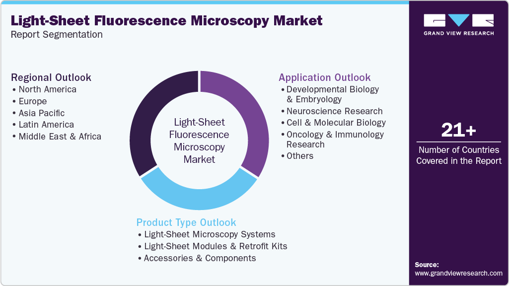
This report forecasts revenue growth at global, regional, and country levels and provides an analysis on industry trends in each of the segments from 2023 to 2035. For the purpose of this study, Grand View Research, Inc. has segmented the global light-sheet fluorescence microscopy market report on the basis of product type, application, and region:
 To learn more about this report, Download Free Sample Report
-
Product Type Outlook (Revenue in USD Million, 2023 - 2035)
-
Light-Sheet Microscopy Systems
-
Light-Sheet Modules & Retrofit Kits
-
Accessories & Components
-
-
Application Outlook (Revenue in USD Million, 2023 - 2035)
-
Developmental Biology & Embryology
-
Neuroscience Research
-
Cell & Molecular Biology
-
Oncology & Immunology Research
-
Others
-
-
Regional Outlook (Revenue in USD Million, 2023 - 2035)
-
North America
-
U.S.
-
Canada
-
Mexico
-
-
Europe
-
UK
-
Germany
-
France
-
Italy
-
Spain
-
Sweden
-
Denmark
-
Norway
-
-
Asia Pacific
-
China
-
India
-
Japan
-
Australia
-
South Korea
-
Thailand
-
-
Latin America
-
Brazil
-
Argentina
-
-
MEA
-
South Africa
-
Saudi Arabia
-
Kuwait
-
UAE
-
-
Research Methodology
The light-sheet fluorescence microscopy market figures in this report are based on a proven research process that combines executive interviews with secondary research from proprietary databases, company filings, and recognized regulatory and institutional sources. Market size is built through value-chain sizing-reconciling supply-side and demand-side estimates-and triangulated with bottom-up and top-down approaches. Every estimate passes multiple levels of expert validation before publication, with each light-sheet fluorescence microscopy segment quantified using the revenue-capture definitions in the table below.
Segment Definition
Product Type
Revenue capture definition
Light-Sheet Microscopy Systems
Complete microscopy platforms designed to generate optical sections of biological samples using sheet-shaped illumination for high-speed 3D imaging with minimal phototoxicity.
Light-Sheet Modules & Retrofit Kits
Specialized illumination and detection modules or upgrade kits integrated into existing microscopes to enable light-sheet imaging capabilities.
Accessories & Components
Supporting hardware such as lasers, cameras, objectives, filters, sample holders, optical components, and software required for light-sheet microscopy operations.
By Application
Revenue capture definition
Developmental Biology & Embryology
Used to visualize embryonic development, organ formation, tissue differentiation, and cellular movements in living organisms.
Neuroscience Research
Enables 3D imaging of brain structures, neuronal networks, synaptic connections, and neural development.
Cell & Molecular Biology
Supports the study of cellular structures, molecular interactions, intracellular processes, and dynamic biological activities.
Oncology & Immunology Research
Used to investigate tumor architecture, cancer progression, immune-cell interactions, and responses to therapeutic treatments.
Others
Includes applications in plant biology, regenerative medicine, tissue engineering, microbiology, and pharmaceutical research.
Estimation Model
Section
Key Summary
1.1 Research Approach
A hybrid research approach combining secondary and primary research methodologies is used to assess the light-sheet fluorescence microscopy market. The assessment considers equipment sales, system shipments, average selling prices, installation volumes, installed base, replacement cycles, and demand across life sciences, biomedical research, pharmaceutical R&D, and academic research applications. Insights from light-sheet microscopy manufacturers, biotechnology and pharmaceutical companies, academic research institutions, microscopy facility managers, distributors, and industry experts are used to validate market estimates. Technological developments in high-speed volumetric imaging, low-phototoxicity illumination, automated sample handling, advanced fluorescence detection, and AI-assisted image reconstruction are also evaluated to understand changes in purchasing patterns and technology adoption.
1.2 Market Estimation Framework
Market size is estimated using three approaches: (1) equipment sales and installation model, (2) installed base and replacement demand model, and (3) key vendor revenue validation model. These approaches are triangulated to estimate the overall market value and develop forecast projections. The framework accounts for differences in system configurations, illumination technologies, imaging resolution, sample compatibility, fluorescence detection capabilities, and application requirements across developmental biology, neuroscience, cell and molecular biology, oncology, and other research applications.
1.2.1 Demand Model
The demand model estimates market demand based on light-sheet microscopy system installations, procurement activity, research infrastructure investments, replacement requirements, and adoption across academic institutions, pharmaceutical companies, biotechnology organizations, and specialized imaging facilities. Demand for complete light-sheet microscopy systems, light-sheet modules and retrofit kits, and accessories and components is considered. Additional factors include increasing research involving organoids, 3D cell cultures, tissue models, whole-organ imaging, embryonic development, and neuronal networks. Investments in advanced microscopy infrastructure, expansion of drug discovery research, and growing demand for long-term live-cell imaging are also evaluated to understand equipment purchasing requirements.
1.2.2 Revenue Model
Market value is calculated based on the number of light-sheet fluorescence microscopy systems and related products sold and their corresponding average selling prices. The model considers variations in system configurations, illumination geometry, laser sources, objective specifications, camera sensitivity, fluorescence detection capabilities, imaging speed, automation, and software integration. Revenues from light-sheet microscopy modules, retrofit kits, accessories, components, installation, maintenance, upgrades, and related software are included where they fall within the defined market scope. Pricing differences between conventional light-sheet systems, lattice light-sheet platforms, selective-plane illumination microscopy (SPIM) systems, and advanced high-throughput imaging platforms are also evaluated.
1.2.3 Vendor Model
Market estimates are validated using revenues, product portfolios, shipment activity, financial disclosures, product launches, and strategic developments of key light-sheet fluorescence microscopy suppliers. The assessment considers manufacturers such as Carl Zeiss AG, Bruker Corporation, Leica Microsystems, Miltenyi Biotec, Intelligent Imaging Innovations (3i), PhaseView, Applied Scientific Instrumentation (ASI), and other relevant suppliers offering light-sheet microscopy systems, modules, and related technologies. Where companies report combined revenues across multiple microscopy or life sciences equipment categories, the light-sheet microscopy contribution is estimated using product-level information, application exposure, product pricing, and other available evidence instead of attributing total company revenue to the market.
1.3 Forecast Development
Forecast projections are developed by analyzing investments in life sciences research, expansion of academic and pharmaceutical research infrastructure, biotechnology R&D expenditure, and increasing adoption of advanced 3D imaging techniques. Demand for organoid research, 3D cell cultures, whole-organ imaging, developmental biology, neuroscience, and preclinical drug discovery is assessed in relation to the need for high-resolution volumetric imaging and reduced phototoxicity. Technological developments in lattice light-sheet microscopy, open-top imaging systems, automated sample handling, high-throughput imaging, AI-enabled image reconstruction, and multimodal fluorescence detection are considered alongside equipment replacement cycles, research funding, laboratory expansion, and the adoption of advanced microscopy platforms across major research regions.
1.4 Segmentation Method
Market segmentation is determined using a weighted assessment of equipment demand, system pricing, installation volumes, application-specific adoption, research expenditure, and end-user procurement patterns. The analysis considers relevant product categories, applications, end users, and geographic regions.
Product-level assessment differentiates light-sheet microscopy systems, light-sheet modules and retrofit kits, and accessories and components based on their functionality, configuration, and contribution to market revenue. Application-level assessment evaluates demand across developmental biology and embryology, neuroscience research, cell and molecular biology, oncology and immunology research, and other applications. End-user analysis considers demand from academic and research institutions, pharmaceutical and biotechnology companies, hospitals and clinical research organizations, and other relevant organizations. Regional estimates account for differences in research funding, biomedical infrastructure, biotechnology investments, technology adoption, and microscopy equipment procurement requirements.
1.5 Scope & Assumptions
The market is measured in USD based on revenues generated from light-sheet fluorescence microscopy products and associated offerings within the defined market scope. Estimates primarily incorporate sales of complete light-sheet fluorescence microscopy systems, including SPIM, lattice light-sheet, and other commercially available light-sheet configurations. Light-sheet modules and retrofit kits, accessories, components, software, installation, maintenance, and upgrade revenues are included only where explicitly covered by the market definition and supported by available data. General-purpose fluorescence microscopes, confocal microscopes, conventional widefield microscopes, standalone optical imaging systems, and unrelated microscopy technologies are excluded unless they meet the defined inclusion criteria. Revenues from research services, contract imaging services, and biological sample analysis are excluded unless explicitly included in the market scope. Regional variations in system pricing, research infrastructure, funding availability, technology adoption, and replacement demand are considered. Revenues from broader microscopy equipment categories are excluded to prevent market overlap and double counting.
Delivered Customizations
This report has been delivered with the following In-depth customizations
Client Request
Customization Delivered
Value Adds
Light-Sheet Microscopy System Pricing & Product Tier Analysis
Comparative assessment of light-sheet microscopy systems across open-top, single-view, dual-view, lattice light-sheet, and selective-plane illumination systems, covering imaging resolution, acquisition speed, sample compatibility, automation, and system capabilities.
Supports price benchmarking, product positioning, technology comparison, and identification of suitable systems across academic, pharmaceutical, and biotechnology research budgets.
Application × Equipment Opportunity Matrix
Mapping of light-sheet microscopy systems and configurations against developmental biology, neuroscience, cell and molecular biology, oncology, immunology, and organoid research applications.
Identifies high-potential applications, unmet imaging requirements, technology gaps, and opportunities for manufacturers to develop application-specific microscopy platforms.
Light-Sheet System & Component Demand Analysis
Assessment of demand for complete microscopy systems, light-sheet modules, retrofit kits, cameras, lasers, objectives, optical components, and image-processing software.
Helps manufacturers identify high-demand product categories, component-level revenue opportunities, and potential cross-selling and aftermarket opportunities.
Research Facility Capacity & Equipment Utilization Analysis
Assessment of light-sheet microscopy installations, equipment utilization, imaging workloads, sample-processing capacity, replacement cycles, and procurement requirements across universities, research institutes, pharmaceutical companies, and biotechnology laboratories.
Supports demand forecasting, capacity planning, installed-base assessment, and identification of opportunities for additional installations, upgrades, and facility expansion.
Advanced 3D Imaging & Drug Discovery Opportunity Assessment
Evaluation of light-sheet microscopy requirements for organoids, 3D cell cultures, whole-organ imaging, live-cell imaging, drug screening, and preclinical toxicity studies, including throughput and automation requirements.
Identifies emerging application opportunities, technology bottlenecks, and commercial growth potential associated with increasing adoption of advanced 3D biological imaging and drug discovery workflows.
About the Author(s)
Medical Devices Research Team
Healthcare · Medical DevicesThis report was authored by the medical devices research team at Grand View Research - comprising two research analysts, one senior research analyst, and one industry expert - with specialized expertise in the medical devices segment of the healthcare industry. All findings are based on proprietary healthcare databases, executive interviews, and regulatory analysis, subject to internal peer review prior to publication.
Last Updated:
Speak to Analyst

Customize this report to your needs - add regions, segments, or data points, with 20% free customization.
Or view our licence options:


ISO 9001:2015 & 27001:2022 Certified
We are GDPR and CCPA compliant! Your transaction & personal information is safe and secure. For more details, please read our privacy policy.


