
- Home
- »
- Medical Devices
- »
-
Phase Contrast Microscopes Market Size Report, 2026-2033GVR Report cover

Phase Contrast Microscopes Market (2026 - 2033)
Size, Share & Trends Analysis Report By Product (Upright Phase Contrast Microscopes, Inverted Phase Contrast Microscopes), By Application (Life Sciences, Material Science), By Region, And Segment Forecasts
Market Size, 2025
$529.8MMarket Estimate, 2026
$562.8MMarket Forecast, 2033
$889.2MCAGR, 2026–2033
6.8%Phase Contrast Microscopes Market Summary
The global phase contrast microscopes market size was valued at USD 529.8 million in 2025 and is projected to grow from USD 562.8 million in 2026 to USD 889.2 million by 2033, at a CAGR of 6.8% from 2026 to 2033. The North America held the largest share of 38.9% of the global market in 2025. This growth is primarily attributed to the growing demand for advanced, non-invasive imaging techniques in life sciences, biotechnology, and pharmaceutical research.
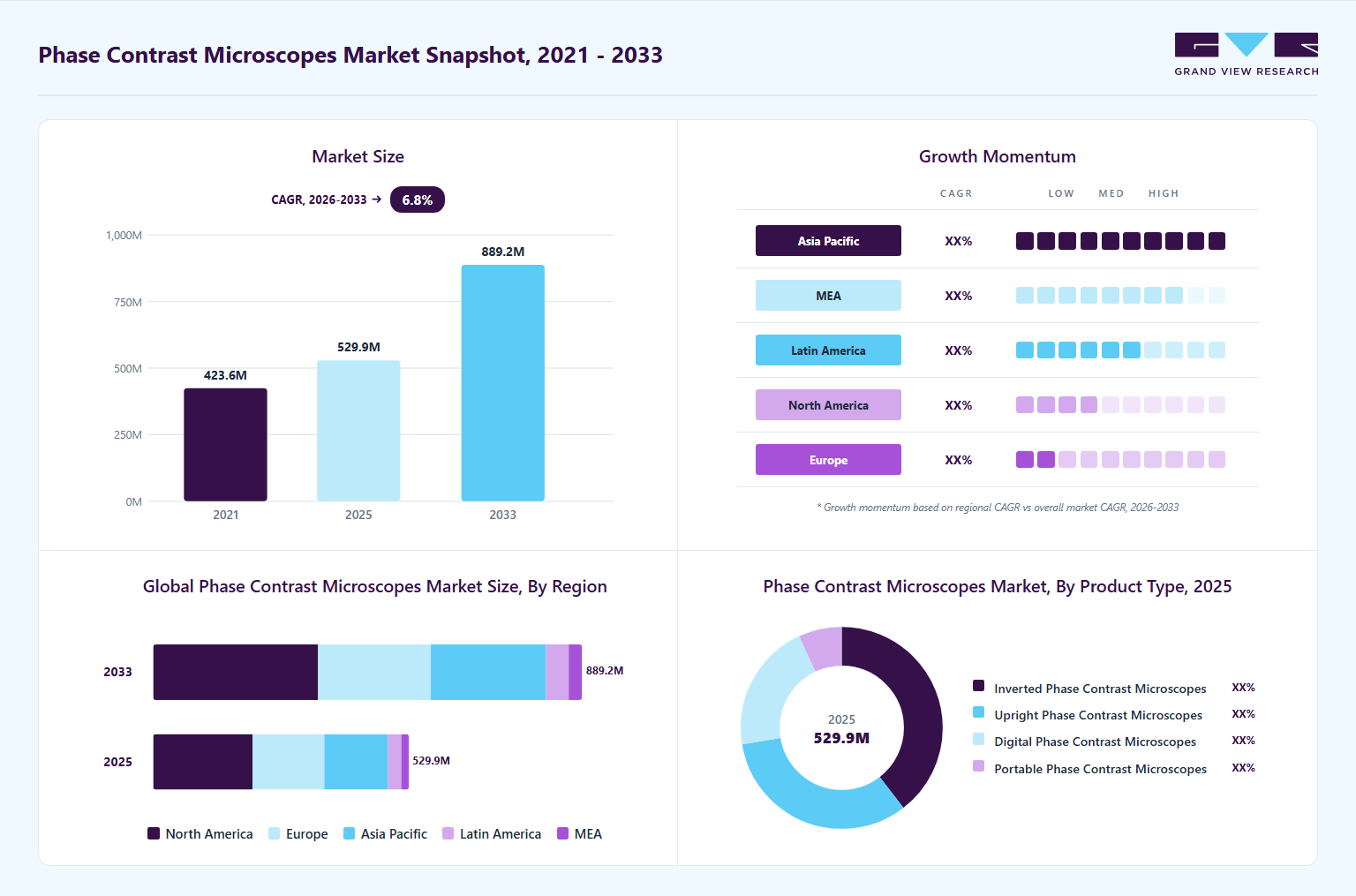 Source: Grand View Research, IR Documents, Primary Interviews, Paid DatabasesTo learn more about this report,
 Download Free Sample Report
 Key Market Trends & Insights
- By product: Inverted phase contrast microscopes segment held the largest market share of 39.5% in 2025.
- By application: Life sciences segment accounted for the largest share of 34.3% in 2025.
Regional Highlights
- Largest regional market: North America (38.9% revenue share, 2025)
- Fastest-growing regional market: Asia Pacific (highest CAGR, 2026-2033)
Market Size & Forecast
- Market size in 2025: USD 529.8 Million
- Estimated market size in 2026: USD 562.8 Million
- Projected market size by 2033: USD 889.2 Million
- CAGR (2026-2033): 6.8%
Three ways to get this report
Buy it, customize it, or ask a question.Ask an Analyst
Analyst response in 1 business day
- AccessReport author, directly
- Reply timeWithin 1 working day
- No sales callDirect answer only
- FormatEmail reply
- CostFree, no cost
Get the Full Report
What the study covers
- FormatsPDF · Excel · Dashboard
- Timeline2026–2033 annual, 2025 base
- Coverage20+ countries, 5 regions
- Companies10+ key players profiled
Customize the Report
20% free customization
- IncludeCountries, segments, points
- TailorTo your product definition
- ExtendDeeper competitor detail
- OrA fully bespoke study
- Turnaround5–10 working days
Moreover, rising investments in biomedical research, precision medicine, and infectious disease studies are further increasing the adoption of phase contrast microscopy across academic institutes, hospitals, and research laboratories.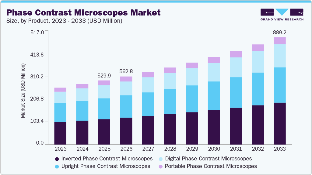 To learn more about this report, Download Free Sample Report
 The section below outlines the key factors driving the growth of the market, highlighting increasing demand from life sciences and biomedical research, rising adoption of live-cell imaging techniques, and expanding applications across biotechnology, pharmaceuticals, clinical diagnostics, and materials science. It also examines the impact of technological advancements such as digital imaging, automation, AI-powered analysis, cloud connectivity, and remote microscopy workflows in improving imaging efficiency, research accuracy, and laboratory productivity, thereby strengthening the adoption of phase contrast microscopes across research and industrial settings.
Growing Life Sciences & Biomedical Research:
Research areas such as cell biology, microbiology, stem cell therapy, cancer research, immunology, regenerative medicine, and pharmaceutical development increasingly require advanced imaging tools capable of observing living cells in their natural state. Phase contrast microscopes are particularly valuable because they allow scientists to visualize transparent and unstained biological specimens with high contrast, eliminating the need for chemical staining that can damage or alter live cells. This capability is critical for long-term live-cell imaging, cell culture monitoring, tissue engineering studies, and drug discovery applications where researchers need to continuously observe cellular behavior, division, migration, and intracellular dynamics in real time. As biotechnology and pharmaceutical companies continue to expand R&D investments, laboratories are increasingly adopting advanced phase contrast imaging systems to improve research efficiency, accuracy, and reproducibility.
Recent technological developments further highlight the growing importance of advanced microscopy techniques in biomedical research. In 2025, researchers published several breakthroughs in live-cell imaging and super-resolution microscopy, demonstrating increasing demand for high-performance imaging systems. For instance, a study published by Nature Photonics in March 2025 introduced a fast three-dimensional live-cell super-resolution imaging method capable of capturing dynamic cellular processes with enhanced speed and resolution, supporting the broader trend toward advanced live-cell microscopy solutions. In addition, in October 2025, researchers at the University of Tsukuba developed an advanced external apodized phase contrast (ExAPC) microscopy technique that enabled high-resolution visualization of cellular organelles without labeling, showcasing the expanding role of phase contrast imaging in organelle and biomolecular research.
Increasing Demand for Live-Cell Imaging:
The increasing demand for live-cell imaging is a major driver for the global phase contrast microscopes industry, as researchers and clinicians increasingly require non-invasive and real-time visualization of living cells without the need for staining or labeling. Phase contrast microscopy is particularly valuable because it allows scientists to observe transparent biological specimens such as live cells, bacteria, tissues, and microorganisms while preserving their natural state and viability. This capability is essential in applications including drug discovery, cancer biology, stem cell research, regenerative medicine, immunotherapy, and clinical diagnostics, where continuous monitoring of cellular behavior, morphology, division, migration, and intracellular interactions is critical.
As pharmaceutical and biotechnology companies increasingly focus on personalized medicine, cell-based therapies, and advanced biologics, the adoption of advanced live-cell imaging systems continues to accelerate, thereby driving demand for phase contrast microscopy technologies.
A November 2025 study published in Nature npj Regenerative Medicine introduced a label-free quantitative phase imaging platform for continuous monitoring of immune cell cultures in bioreactors. Technology enables real-time, non-destructive observation of T-cell cultures and CAR-T cells without staining or disrupting the sterile culture environment. This development highlights the growing importance of phase contrast and quantitative phase imaging technologies in cell therapy manufacturing, immunotherapy research, and regenerative medicine, thereby supporting increasing demand for advanced live-cell imaging systems in the market.
The pharmaceutical sector is also increasingly utilizing live-cell imaging for drug screening and cytotoxicity analysis.
Case Study: Queen Mary University of London’s FRAP-SR Innovation Advancing Live-Cell Imaging
Background:
In August 2025, researchers at Queen Mary University of London, in collaboration with Carl Zeiss and Rapp OptoElectronics, developed an advanced live-cell imaging technique called FRAP-SR (Fluorescence Recovery After Photobleaching in Super-Resolution Regime). The innovation combined Lattice Structured Illumination Microscopy (diSIM/SIM²) with FRAP technology to achieve ultra-high-resolution imaging of living cells while minimizing phototoxicity and cellular damage.
Development:
The FRAP-SR technique enabled researchers to visualize cellular structures at a resolution of approximately 60 nanometres, allowing detailed observation of nanoscale biological processes in living cells. Using the technology, researchers studied the DNA repair protein 53BP1 and discovered complex subcompartments within DNA repair condensates that exhibited varying protein mobility and functional specialization. The study demonstrated the capability of advanced microscopy systems to capture highly dynamic intracellular activities in real time without affecting cell viability.
The research utilized the ZEISS Elyra 7 platform integrated with advanced FRAP capabilities, highlighting the increasing adoption of technologically advanced imaging systems in biomedical laboratories and research institutions.
Market Impact:
This development highlights the growing importance of advanced live-cell imaging technologies in life sciences and biomedical research, which is significantly driving demand for phase contrast and super-resolution microscopy systems. Researchers increasingly require non-invasive imaging tools capable of observing live cells, DNA repair mechanisms, chromosome dynamics, and cellular interactions with high precision and minimal disturbance. The innovation also supports expanding applications in cancer research, regenerative medicine, molecular biology, and drug discovery.
Furthermore, the ability of FRAP-SR technology to support live-cell drug screening and personalized medicine research is expected to accelerate investments in advanced microscopy platforms across the pharmaceutical and biotechnology industries. The study also reflects the broader industry trend toward integrating high-resolution optics, fluorescence imaging, and computational microscopy technologies to improve research efficiency and biological analysis.
Strategic Significance:
The project was supported by a USD 2.46 million grant from UK Research and Innovation and facilitated the establishment of the Center for Cell Dynamics at Queen Mary University of London. The case demonstrates how collaborations between academic institutions and microscopy manufacturers are promoting innovation and accelerating the commercialization of next-generation microscopy technologies, thereby contributing to the growth of the global phase contrast microscopes industry.
Key opinion:
"Our FRAP-SR approach enables us to visualize structures as small as 60 nanometres within living cells - a scale previously inaccessible for dynamic studies without causing significant cellular stress. "This resolution, 2000 times smaller than the width of a human hair, allows us to probe the nanoscale organization and behavior of cellular components in real-time." -team lead
Growing Adoption in Academic & Educational Institutions:
Universities, medical colleges, life science institutes, and research laboratories are increasingly incorporating advanced microscopy systems into their teaching, training, and research programs to support modern biological and biomedical education. Phase contrast microscopes are particularly valuable in academic environments because they allow students and researchers to observe live, transparent, and unstained specimens such as cells, bacteria, protozoa, and tissue cultures with high clarity and minimal sample preparation. Their ease of operation, cost-effectiveness compared to highly advanced imaging systems, and ability to provide real-time visualization of biological processes make them highly suitable for laboratory training and experimental research.
In addition, the rising emphasis on STEM education, biotechnology courses, regenerative medicine studies, and cell biology research is encouraging institutions globally to upgrade microscopy infrastructure and establish advanced imaging facilities. Increasing government and institutional funding for scientific research, coupled with collaborations between universities and microscope manufacturers, is further accelerating adoption across academic settings.
Moreover, in September 2025, Monash University partnered with Nikon to establish Australia’s first Nikon Center of Excellence, focused on next-generation light microscopy research and education. The initiative strengthens the university’s microscopy infrastructure by adding advanced imaging systems, including Nikon confocal and widefield microscopes, across multiple research facilities. The center is also expected to serve as a training hub to educate students and researchers on modern microscopy techniques and imaging technologies. This development highlights the growing adoption of advanced microscopy systems in academic institutions, supporting the demand for phase contrast and other optical microscopes in education and life sciences research.
“The establishment of the new Nikon Center of Excellence is an exciting milestone that strengthens our position as one of Australia’s leading optical microscopy facilities. These state-of-the-art microscopes will provide faster and higher-resolution imaging for hundreds of researchers across four sites within Monash Medicine, Nursing and Health Sciences, and our partner research institutes.”- Manager of Monash Micro Imaging
Similarly, in January 2025, researchers at the University of Strathclyde developed the world’s first fully 3D-printed microscope, designed specifically to improve affordable access to microscopy tools for students and researchers. The low-cost system demonstrates how educational institutions are increasingly focusing on accessible microscopy technologies to enhance practical scientific learning and laboratory training.
“In under three hours, you can transform a design, freely available on the internet, into a fully functional optical microscope. “This opens the doors to democratized access, rapid prototyping, and bespoke design of microscopes and optics at a fraction of the price of traditional microscopes.” “It could help scientists and medics in low-income countries around the world, as well as enabling students to learn more about science through an accessible, cheap kit.”- Postdoctoral research associate, who created the device with Professor Gail McConnell
Expansion of Nanotechnology & Materials Science Research:
The expansion of nanotechnology and materials science research is emerging as a major driver for the market, as researchers increasingly require advanced imaging systems capable of analyzing nanoscale structures, semiconductor materials, thin films, and microstructural properties with high precision. Phase imaging techniques are gaining importance because they allow scientists to observe transparent or weakly scattering materials without staining or destructive preparation methods.
In nanotechnology and semiconductor research, these microscopes help researchers study nanoparticle interactions, crystal defects, surface morphology, electromagnetic fields, and structural transformations at micro- and nanoscale resolutions. The rapid growth of industries such as electronics, quantum computing, energy storage, and advanced materials is further accelerating demand for high-resolution phase imaging systems that support real-time and non-destructive analysis.
Moreover, recent advancements in differential phase-contrast STEM imaging are enabling researchers to visualize electromagnetic fields and semiconductor device structures at nanometer and sub-angstrom scales. These innovations are expected to support future developments in spintronics, quantum devices, and nanoelectronics, further strengthening demand for phase contrast and phase imaging globally.
Case Study: Modulated Optically Computed Phase Microscopy (M-OCPM) for Imaging Cells and Nanoparticles
Background:
The increasing use of nanoparticles in drug delivery systems has created a strong demand for advanced imaging technologies capable of studying interactions between nanoparticles and living cells. Conventional microscopy techniques often face limitations in simultaneously achieving high spatial resolution and high phase sensitivity, which are essential for observing nanoscale biological processes. To address this challenge, researchers developed a novel imaging technology called Modulated Optically Computed Phase Microscopy (M-OCPM), published in Scientific Reports in January 2025 by researchers.
Objective:
The study aimed to develop a high-resolution, label-free quantitative phase imaging technology capable of visualizing nanoparticle-cell interactions with nanometer-scale sensitivity. The goal was to overcome the traditional tradeoff between imaging sensitivity and spatial resolution commonly observed in phase microscopy systems.
Technology Innovation:
M-OCPM combines low-coherence interferometry with optical computation to improve imaging performance. The key innovation involves optical Fourier transform computation and temporal modulation of interferometric signals, enabling the system to bypass the conventional sensitivity-resolution limitation without relying on mechanical scanning. The technology achieved:
-
High spatial resolution of approximately 250 nm
-
Nanometer-scale displacement sensitivity
-
Label-free imaging capability for live cells and nanoparticles
-
Enhanced visualization of subtle refractive index changes caused by nanoparticles
Key Findings:
Researchers successfully imaged cultured cells together with nanoparticles and differentiated nanoparticles adhered to cells from those suspended in culture medium. The system demonstrated the ability to:
-
Detect nanoscale structural changes in biological samples
-
Resolve adjacent nanoparticles individually
-
Observe dynamic cell-NP interactions in real time
-
Improve visualization of transparent biological specimens without staining
The study validated M-OCPM as a powerful tool for analyzing nanoparticle-based drug delivery systems and nano-biological interfaces.
Market Impact:
This development highlights the growing importance of advanced phase contrast and quantitative phase imaging technologies in nanotechnology, materials science, and biomedical research. As pharmaceutical companies increasingly invest in nanoparticle-based therapeutics, vaccine delivery systems, and precision medicine, demand for high-resolution microscopy solutions is rising rapidly.
Technology is particularly relevant for:
-
Nanomedicine and targeted drug delivery research
-
Semiconductor and nanoscale materials inspection
-
Biomedical and life science imaging
-
Preclinical pharmaceutical R&D
-
Cell biology and regenerative medicine research
Strategic Importance for the Market:
The M-OCPM breakthrough demonstrates how next-generation phase imaging systems are evolving beyond traditional biological microscopy into advanced nanotechnology and materials characterization applications. Such innovations are expected to accelerate the adoption of sophisticated phase contrast microscopy platforms across research institutes, pharmaceutical companies, semiconductor laboratories, and nanotechnology centers globally. The increasing requirement for non-destructive, real-time, high-sensitivity imaging is likely to remain a significant growth driver for the global market.
Shift Toward Digital & Automated Microscopy Systems:
The shift toward digital and automated microscopy systems is becoming a major growth driver for the market as laboratories, hospitals, pharmaceutical companies, and research institutions increasingly seek faster, more accurate, and user-friendly imaging solutions. Traditional microscopy methods often require manual adjustments, physical observation through eyepieces, and on-site analysis, which can limit workflow efficiency and data sharing. In contrast, modern digital phase contrast microscopes integrate automated focusing, motorized stage movement, AI-powered image analysis, cloud connectivity, and remote access capabilities, enabling researchers and clinicians to conduct real-time observation, automated data capture, and collaborative analysis from different locations. These advanced systems significantly improve productivity, reduce human error, support high-throughput workflows, and enable seamless integration with laboratory information systems. The growing adoption of telepathology, remote diagnostics, and digital healthcare infrastructure is further accelerating demand for connected microscopy platforms across life sciences, pathology, biotechnology, and materials science applications.
In September 2025, Olympus Corporation launched cloud-based OLYSENSE CAD/AI applications in Europe to support connected diagnostic workflows and AI-assisted analysis, reflecting the broader industry movement toward intelligent and digitally integrated imaging ecosystems.
“Cloud-based and AI-powered endoscopy solutions are transforming clinical practice. By enabling real-time decision support and standardized assessments, they help clinicians diagnose earlier and more accurately, streamline workflows, and improve patient outcomes. Endoscopy on the cloud opens the door for continuous innovation and broader access to advanced care.”
-Professor in Gastroenterology from Humanitas University, Italy
“With OLYSENSE, we are taking a meaningful step forward in helping patients receive earlier, more accurate diagnoses. Our goal is simple: to give clinicians the advanced tools that make it easier to find and assess challenging lesions early, make confident, informed decisions, and provide their patients with the best possible care.”
- Corporate Officer, Head of Gastrointestinal Solutions Division, Olympus Corporation
Furthermore, in March 2025, Nikon Corporation Healthcare Business Unit introduced Version 1.4 of its ECLIPSE Ui digital imaging microscope, featuring enhanced remote operation functionality, simultaneous comparison of up to 10 specimen images, and network-based viewing capabilities to improve pathology workflows and telemedicine support.
Market Characteristics & Concentration
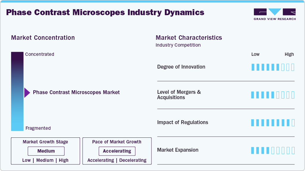
The chart below represents the relationship between industry concentration, industry characteristics, and industry participants. There is a moderate degree of innovation, a low level of merger & acquisition activities, high impact of regulations, and moderate expansion of industry.
The industry is experiencing a moderate degree of innovation. The ZEISS Versa X-ray Microscopes represent a significant innovation in high-resolution 3D imaging, extending the capabilities of conventional microscopy into the realm of non-destructive volumetric analysis. Developed by ZEISS, the Versa X-ray Microscopy (XRM) platform enables researchers to visualize internal structures of materials and biological samples in three dimensions at submicron resolution without sectioning or destroying the specimen. This innovation is particularly important for advanced research fields such as materials science, semiconductor inspection, energy storage (battery research), and biomedical imaging, where understanding internal microstructures is critical.
 To learn more about this report, Download Free Sample Report
 Several key players are actively engaging in mergers & acquisitionsto promote growth & innovation and improve their competitiveness by combining the expertise & efforts of different organizations. For instance, the acquisition of Phasefocus Holdings Limited by Bruker Corporation in March 2024 represents a significant development in the advanced cell imaging and microscopy space, further strengthening its capabilities in label-free and quantitative imaging technologies. The acquisition also reflects the growing industry trend toward advanced phase-based and quantitative imaging techniques, which extend beyond traditional phase contrast microscopy by combining optical physics with powerful image reconstruction algorithms.
Regulations play a crucial role in driving the industry as they enforce strict standards for safety, hygiene, waste management, and operational efficiency in healthcare environments.
-
In medical and clinical applications, phase contrast microscopes fall under medical device regulations in many regions. For instance, in Europe, these instruments must comply with the EU Medical Device Regulation (MDR 2017/745) if used for diagnostic purposes, requiring CE marking, clinical evaluation, and strict quality management system compliance.
-
For laboratory and research use, these microscopes are generally governed by organizations such as the International Organization for Standardization that define key quality and performance benchmarks, including optical performance, calibration accuracy, and safety standards under ISO frameworks (such as ISO 9001 for quality management and ISO 13485 for medical device manufacturing).
KA Imaging’s global expansion highlights the accelerating adoption of spectral X-ray imaging technologies across clinical and research settings. In April 2026, the company strengthened its presence in the Asia-Pacific region by obtaining an Irradiating Apparatus License in Hong Kong for its Reveal Mobi Pro and Mobi Lite mobile X-ray systems, while also receiving regulatory clearance in Australia for commercial deployment of the Reveal Mobi Pro. This expansion reflects increasing clinical demand for advanced mobile imaging solutions that enhance diagnostic information without increasing operational complexity, particularly in bedside and emergency care environments.
“By bringing together phase-contrast imaging with inCiTe and high-resolution amorphous selenium technology with BrillianSe, we’re helping advance research and science,”
-Vice President of NDT Sales & Business Development.
Product Insights
The inverted phase contrast microscopes segment held the largest share of 39.5% in 2025 due to its critical role in live-cell imaging and advanced biological research, where observing cells in their natural, unaltered state is essential. These microscopes place the objective lens below the specimen, making them ideal for examining cells in culture dishes, Petri dishes, and multi-well plate formats widely used in modern laboratories. For instance, the Nikon Eclipse Ti2 / Ti-S is a high-end research inverted microscope widely used in live-cell imaging and phase contrast applications. It supports modular configurations, motorized stages, and digital imaging systems for advanced biological research.
The digital phase contrast microscopes segment is expected to grow at the fastest CAGR during the forecast period. This growth is attributed to the rising demand for high-quality live-cell imaging and real-time visualization, especially in life sciences, where researchers need to observe transparent biological samples without staining or damaging them. Digital phase contrast systems enhance traditional phase contrast microscopy by integrating high-resolution cameras, image processing software, and digital display systems, allowing users to capture, store, and analyze images more efficiently than conventional eyepiece-based microscopes. For instance, the Axiovert 5 digital is an all-in-one cell imaging system that integrates phase contrast imaging with digital image capture, AI-based cell analysis, and automated workflows for life science applications.
Application Insights
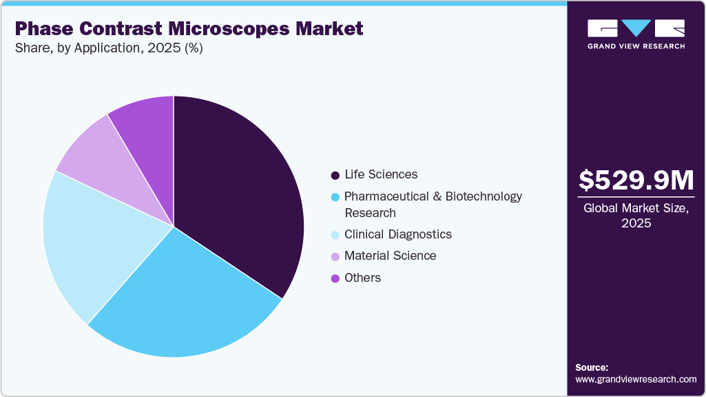
Based on application, life sciences accounted for the largest share of 34.3% in 2025 due to the expansion of biomedical and pharmaceutical R&D activities, where phase contrast microscopes are widely used in drug discovery, toxicity testing, and disease modeling. For instance, in cancer research, they help monitor tumor cell behavior and response to treatment at the cellular level, while in microbiology, they are used for studying bacteria and protozoa without altering their natural state. These microscopes are uniquely suited for observing live, unstained biological samples such as cells, tissues, microorganisms, and intracellular structures.
 To learn more about this report, Download Free Sample Report
 The pharmaceutical & biotechnology research segment is expected to grow at the fastest CAGR over the forecast period as these industries rely heavily on detailed observation of living cells and transparent biological samples during drug discovery, development, and testing processes. The expansion of drug discovery and preclinical research pipelines, particularly in oncology, immunology, and infectious diseases. Pharmaceutical companies increasingly use phase contrast microscopes to monitor morphological changes in cells during compound screening, helping identify promising drug candidates faster and more efficiently.
Regional Insights
North America phase contrast microscope market held the largest revenue share of 38.9% in 2025, owing to the region’s strong research infrastructure, high technological adoption, and diverse industrial and scientific applications that together create sustained demand for advanced optical imaging solutions. The region is led by the U.S. with its substantial investments in R&D, well-funded academic institutions, and prominent biomedical and life sciences research facilities, which require high-precision visualization tools for cellular biology, materials science, and diagnostic workflows, which bolsters the uptake of microscopes in laboratories and clinical environments.
 To learn more about this report, Download Free Sample Report
 U.S. Phase Contrast Microscope Market Trends
The phase contrast microscope market in the U.S.is experiencing significant growth, driven by the new developments and product launches. For instance, in May 2024, Nikon introduced the SI-PH Phase Condenser accessory for its ECLIPSE Si biological microscope, representing a targeted advancement in phase contrast microscopy designed to enhance efficiency and usability in research and educational environments. By integrating this accessory with the ECLIPSE Si platform, Nikon aims to streamline workflow by reducing the need for frequent adjustments and improving image quality consistency during long-duration microscopic examinations.
Asia Pacific Phase Contrast Microscope Market Trends
The phase contrast microscope market in Asia Pacific is expected to grow at the fastest CAGR during the forecast period. This is due to driven by rapid adoption across research, healthcare, agriculture, and educational sectors. Rapid industrialization and expansion of manufacturing hubs, particularly in electronics, semiconductors, and precision engineering, have increased the demand for high-precision visual inspection tools used in quality control, defect analysis, and micro-assembly tasks. This is especially pronounced in countries such as China and Japan, where large automotive sectors prioritize reliable stereo imaging for detailed component inspection.
Japan phase contrast microscope marketheld a significant revenue share in 2025. The launch of the Hitachi High-Tech Miniscope TM4000PlusIII (2024) supports the growth of the market by strengthening the broader ecosystem of advanced, compact, and digital microscopy solutions used in research and industrial laboratories. These innovations encourage broader adoption of complementary optical techniques such as phase contrast microscopy in laboratories that require live-cell imaging and non-invasive specimen observation.
The phase contrast microscope market in India is driven by the presence and growth of microscope manufacturers in the country, as highlighted in the ESAW India article published in August 2025 is a significant driver for the microscope market in the country, as it boosts domestic availability, affordability, and technological relevance of these instruments. Indian manufacturers now produce a wide range of microscopes, including microscopes that offer three-dimensional visualization for quality control, dissection, and industrial inspection, meeting the needs of education, research, healthcare, and industrial labs across India.
Europe Phase Contrast Microscope Market Trends
The phase contrast microscope market inEurope is expected to witness high growth owing to the strong research activity, industrial demand, and technological innovation that together support strong adoption across scientific, healthcare, and quality-control applications. Europe’s well-established life sciences and materials research infrastructure, supported by significant public and private R&D funding (including programs such as Horizon Europe), fuels demand for high-precision imaging tools microscopes are essential for detailed 3D visualization in biological, cellular, and materials studies.
Germany phase contrast microscope marketheld a significant share in the European region, owing to the new product developments and innovative offerings. For instance, in March 2024, UC Enuity represented the next generation of ultramicrotomes, designed to significantly enhance laboratory productivity by combining precision engineering with advanced automation. Its automated setup functions streamline traditionally time-consuming preparation steps, reducing operator dependency and minimizing setup errors while ensuring consistently high-quality ultrathin sections.
James O'Brien, Vice President of Life Sciences at Leica Microsystems, said:
"UC Enuity helps to achieve precise and high-quality results consistently by automating critical steps in sample preparation. This automation makes this technology accessible and embodies our unwavering dedication to empowering the scientific community to unlock new discoveries. This built-to-last ultramicrotome has been designed with the future in mind. Researchers will be able to adapt to their ultramicrotome as they expand their capabilities and maximize their scientific potential."
Latin America Phase Contrast Microscope Market Trends
The phase contrast microscope market inLatin America is expected to register considerable growth over the forecast period, owing to the expanding investments in healthcare, research, and industrial infrastructure, which are gradually improving the accessibility and adoption of microscopy equipment across the region. Countries such as Brazil and Mexico are notable for their ongoing enhancements in healthcare and diagnostic capabilities, which boost demand for tools such as microscopes in clinical laboratories and medical research institutions.
The Brazil phase contrast microscope marketheld the largest regional market share in 2025, driven by the launch of the Brazil-China Joint Synchrotron Science Laboratory at CNPEM in July 2025. The collaboration enhances Brazil’s access to international expertise, funding, and large-scale research projects, leading to increased demand for advanced optical microscopes across national laboratories, universities, and affiliated research centers.
Andrea Brito Latgé, Secretary of Strategic Policies and Programs at MCTI, said:
“We are very pleased with this agreement between CNPEM and IHEP, which strengthens a historic and strategic relationship between Brazil and China. This cooperation, especially in the field of synchrotron light sources, allows the exchange of experiences between laboratories with complementary expertise. There is a mutual desire to reinforce South-South cooperation, accelerating joint project development and involving young researchers, which is essential for advancing science, technology, and innovation in our countries,”
Middle East & Africa Phase Contrast Microscope Market Trends
The phase contrast microscope market inthe Middle East & Africa (MEA) is driven by the growing need for high-precision imaging technologies across multiple sectors. Governments, particularly in countries such as Saudi Arabia and the UAE, are prioritizing the modernization of research laboratories, medical facilities, and academic institutions by funding advanced equipment and promoting innovation as part of broader economic diversification initiatives such as Saudi Vision 2030, which accelerates the adoption of cutting-edge microscopy tools in clinical, research, and industrial settings.
The UAE phase contrast microscope market is driven by increasing visibility, availability, and applicability of advanced optical inspection tools across multiple sectors. The showcasing of advanced optical instruments, including professional-grade phase contrast and digital microscopes at ARABLAB 2024 (held at the Dubai World Trade Center), acts as a key growth catalyst for the UAE market by strengthening product visibility, technology adoption, and cross-border scientific collaboration.
Key Phase Contrast Microscope Companies Insights
The phase contrast microscope market is fragmented, with the presence of many country-level providers.The market players undertake several strategic initiatives, such as partnerships & collaborations, product launches, mergers & acquisitions, and geographical expansion to maintain their position and grow in the market.
Key Phase Contrast Microscopes Companies:
The following key companies have been profiled for this study on the phase contrast microscope market.
- ZEISS
- Olympus
- Thermo Fisher Scientific India Pvt Ltd.
- LW Scientific
- Vision Engineering Ltd
- Meiji Techno America
- EVIDENT
- Leica Microsystems
Recent Developments
- In October 2024, KA Imaging launched a configurable micro-CT system, highlighting a major advancement in the phase contrast microscopes and X-ray imaging market by integrating both phase contrast and spectral imaging technologies into a single modular platform. The incite 2.0 3D X-ray Microscope is designed for industrial and research applications and enables high-resolution, non-destructive imaging of complex materials by combining propagation-based phase contrast with dual-energy spectral imaging.
“With inCiTe 2.0, we’re addressing the growing need for precise, high-quality imaging that can deliver detailed insights across a wide range of industries,” -President and CEO of KA Imaging.
“This system’s ability to combine phase contrast and spectral imaging makes it an invaluable tool for both research and industrial applications, and it serves as a promising alternative to SEM in many cases,” -CTO
- In January 2023, ZEISS introduced an all-in-one cell imaging system, Axiovert 5 digital, representing a significant advancement in the market, reflecting the shift toward intelligent, automated, and all-in-one cell imaging systems. The system integrates artificial intelligence (AI) and fully automated functions to simplify routine laboratory workflows and improve imaging consistency.
Phase Contrast Microscopes Market Report Scope
Report Attribute
Details
Market size in 2025
USD 529.8 million
Estimated market size in 2026
USD 562.8 million
Projected market size by 2033
USD 889.2 million
Growth rate
CAGR of 6.8% from 2026 to 2033
Actual data
2021 - 2025
Forecast period
2026 - 2033
Quantitative units
Revenue in USD million and CAGR from 2026 to 2033
Report coverage
Revenue forecast, company ranking, competitive landscape, growth factors, and trends
Segments covered
Product, application, region
Regional scope
North America; Europe; Asia Pacific; Latin America; MEA
Country scope
U.S.; Canada; Mexico; Germany; UK; France; Italy; Spain; Norway; Denmark; Sweden; China; Japan; India; South Korea; Australia; Thailand; Brazil; Argentina; Saudi Arabia; South Africa; UAE; Kuwait
Key companies profiled
ZEISS; Olympus; Thermo Fisher Scientific India Pvt Ltd.; LW Scientific; Vision Engineering Ltd; Meiji Techno America; EVIDENT; Leica Microsystems
Customization scope
Free report customization (equivalent up to 8 analysts working days) with purchase. Addition or alteration to country, regional & segment scope.
Pricing and purchase options
Avail customized purchase options to meet your exact research needs. Explore purchase options
Global Phase Contrast Microscopes Market Report Segmentation
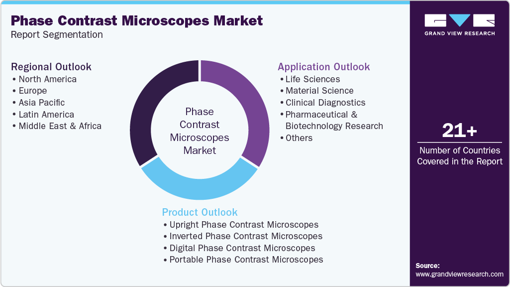
This report forecasts revenue growth at global, regional, and country levels and provides an analysis of the latest industry trends in each of the sub-segments from 2021 to 2033. For this study, Grand View Research has segmented the global phase contrast microscopes market report based on product, application, and region.
 To learn more about this report, Download Free Sample Report
 -
Product Outlook (Revenue, USD Million, 2021 - 2033)
-
Upright Phase Contrast Microscopes
-
Inverted Phase Contrast Microscopes
-
Digital Phase Contrast Microscopes
-
Portable Phase Contrast Microscopes
-
-
Application Outlook (Revenue, USD Million, 2021 - 2033)
-
Life Sciences
-
Material Science
-
Clinical Diagnostics
-
Pharmaceutical & Biotechnology Research
-
Others
-
-
Regional Outlook (Revenue, USD Million, 2021 - 2033)
-
North America
-
U.S.
-
Canada
-
Mexico
-
-
Europe
-
UK
-
Germany
-
France
-
Italy
-
Spain
-
Denmark
-
Sweden
-
Norway
-
-
Asia Pacific
-
China
-
Japan
-
India
-
South Korea
-
Australia
-
Thailand
-
-
Latin America
-
Brazil
-
Argentina
-
-
Middle East and Africa (MEA)
-
South Africa
-
Saudi Arabia
-
UAE
-
Kuwait
-
-
Frequently Asked Questions About This Report
The global phase contrast microscopes market size was valued at USD 529.8 million in 2025 and is estimated at USD 562.8 million for 2026.
The global phase contrast microscopes market is expected to grow at a CAGR of 6.8% from 2026 to 2033, reaching USD 889.2 million by 2033.
Key players in the market include ZEISS, Olympus, Thermo Fisher Scientific India Pvt Ltd., LW Scientific, Vision Engineering Ltd, Meiji Techno America, EVIDENT, Leica Microsystems.
This growth is primarily attributed to the growing demand for advanced, non-invasive imaging techniques in life sciences, biotechnology, and pharmaceutical research.
The inverted phase contrast microscopes segment led with a 39.5% revenue share in 2025, while the digital phase contrast microscopes segment is expected to grow at the fastest CAGR during the forecast period.
The life sciences segment held the largest revenue share of 34.3% in 2025, while the pharmaceutical & biotechnology research segment is expected to grow at the fastest CAGR.
North America dominated the market with a 38.9% revenue share in 2025.
Asia Pacific is the fastest-growing region over the forecast period.
About the Author(s)
Medical Devices Research Team
Healthcare · Medical DevicesThis report was authored by the medical devices research team at Grand View Research - comprising two research analysts, one senior research analyst, and one industry expert - with specialized expertise in the medical devices segment of the healthcare industry. All findings are based on proprietary healthcare databases, executive interviews, and regulatory analysis, subject to internal peer review prior to publication.
Last Updated:
Speak to Analyst

Customize this report to your needs - add regions, segments, or data points, with 20% free customization.
Or view our licence options:


ISO 9001:2015 & 27001:2022 Certified
We are GDPR and CCPA compliant! Your transaction & personal information is safe and secure. For more details, please read our privacy policy.


