- Home
- »
- Medical Devices
- »
-
Confocal Scanning Microscopes Market Size Report, 2033GVR Report cover
![Confocal Scanning Microscopes Market Size, Share & Trends Report]()
Confocal Scanning Microscopes Market (2026 - 2033) Size, Share & Trends Analysis Report By Product Type (Laser Scanning Confocal Microscopes, Spinning Disk Confocal Microscopes), By Application (Life Sciences, Material Science), By Region, And Segment Forecasts
- Report ID: GVR-4-68040-932-1
- Number of Report Pages: 150
- Format: PDF
- Historical Range: 2021 - 2025
- Forecast Period: 2026 - 2033
- Industry: Healthcare
- Report Summary
- Table of Contents
- Segmentation
- Methodology
- Download FREE Sample
-
 Download Sample Report
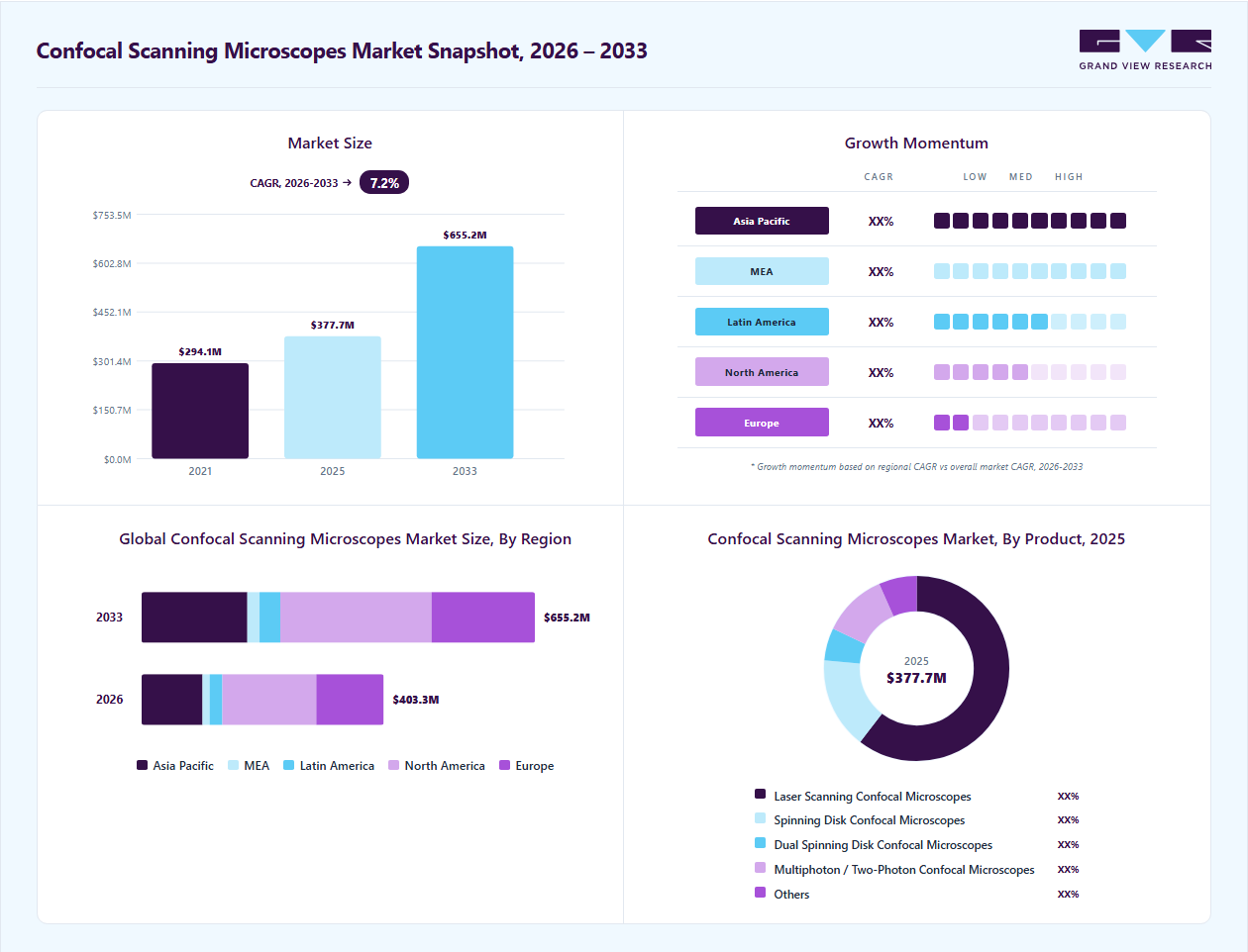
Market Size, 2025$377.7MMarket Estimate, 2026$403.3MMarket Forecast, 2033$655.2MCAGR, 2026 - 20337.2%Confocal Scanning Microscopes Market Summary
The global confocal scanning microscopes market size was valued at USD 377.7 million in 2025 and is projected to grow from USD 403.3 million in 2026 to USD 655.2 million by 2033, at a CAGR of 7.2% from 2026 to 2033. North America market dominated the global market in 2025 and accounted for the largest revenue share of 38.9%. This growth is primarily attributed to the growing demand for advanced imaging technologies in life sciences, clinical diagnostics, and pharmaceutical research.
Key Market Trends & Insights
- In terms of product type segment, the laser scanning confocal microscopes segment held the largest revenue share in 2025.
- Based on application, life science accounted for the largest share of 32.5% in 2025
Regional Highlights
- Largest regional market: North America (38.9% revenue share, 2025)
- Fastest-growing regional market: Asia Pacific (highest CAGR, 2026-2033)
- U.S. market is anticipated to register the fastest growth rate during the forecast period
Market Size & Forecast
- 2025 Market Size: USD 377.7 Million
- 2026 Market Size: USD 403.3 Million
- 2033 Projected Market Size: USD 655.2 Million
- CAGR (2026-2033): 7.2%
In addition, the rapid expansion of biotechnology and pharmaceutical industries, coupled with increasing investments in drug discovery and personalized medicine, is significantly supporting market growth, as researchers require detailed cellular visualization for understanding disease mechanisms and evaluating therapeutic efficacy.")
The section below highlights the key drivers of the confocal scanning microscopes market, including rising demand for high-resolution imaging in life sciences and biomedical research, increasing adoption of live-cell imaging, and expanding applications in biotechnology, pharmaceuticals, diagnostics, and material sciences. It also covers technological advancements such as AI-powered imaging, automation, digital microscopy, and cloud connectivity that are enhancing research efficiency and supporting wider market adoption.
Case Study: Use of Organoids and Confocal Microscopy in Drug Discovery and Personalized Medicine
Background
Traditional preclinical testing methods, including animal models and two-dimensional cell cultures, often fail to accurately predict how drug candidates will behave in humans. This limitation contributes to the high failure rate of drug candidates during clinical trials, increasing development costs and delaying the introduction of new therapies. To overcome these challenges, pharmaceutical and biotechnology companies are increasingly adopting advanced biological models such as organoids, which closely mimic human tissue structure and function.
Objective
The objective of this initiative was to improve drug safety and efficacy prediction through the use of organoids and advanced imaging technologies such as confocal microscopy. The approach aimed to provide more human-relevant data during preclinical testing, reduce dependence on animal models, and support personalized medicine applications.
Technology and Approach
Charles River Laboratories utilized lab-grown organoids, miniature three-dimensional tissue cultures derived from stem cells to replicate human organ behavior in vitro. These organoids were analyzed using high-resolution confocal microscopy, enabling researchers to visualize cellular structures, tissue organization, and molecular interactions in detail.
At the company’s Leiden facility in the Netherlands, researchers developed intestinal organoids containing epithelial cell types such as goblet cells, enterocytes, enteroendocrine cells, and Paneth cells. Confocal microscopy played a critical role in capturing detailed 3D images of these organoids and evaluating cellular responses to antiviral drug candidates.
Key Findings
Researchers revisited three antiviral drug candidates that had previously failed during early-stage clinical trials despite showing positive results in conventional preclinical studies.
-
In two of the three cases, organoid models identified toxicity and efficacy issues that had not been detected in traditional cell cultures or animal testing.
-
One drug demonstrated significantly higher toxicity in intestinal organoids.
-
Another showed reduced therapeutic efficacy along with unexpected toxicity risks.
-
A third compound initially appeared effective; however, detailed confocal imaging revealed that the drug only temporarily inhibited viral replication instead of fully suppressing the virus.
These findings closely aligned with the actual failures observed during human clinical trials, demonstrating the predictive capability of organoid-based testing.
Additional Development in Toxicology Testing
Charles River Laboratories also collaborated with biotechnology company MatTek Corporation to develop advanced lung organotypic models for inhalation toxicology testing. Scientists at the company’s Edinburgh site used human airway epithelial models cultured at the air-liquid interface to assess the inhalation risks of chemicals.
In collaboration with Syngenta, researchers applied these models to evaluate the fungicide chlorothalonil. The study became the first OECD case study where regulatory decisions were supported using lung organoid data instead of traditional animal testing methods.
Role of Confocal Microscopy
Confocal microscopy was essential in this research due to its ability to provide:
-
High-resolution three-dimensional imaging
-
Enhanced visualization of cellular morphology
-
Real-time live-cell imaging
-
Improved analysis of tissue organization and molecular interactions
Technology enabled researchers to accurately monitor organoid behavior, assess drug responses, and identify toxicity signals at the cellular level.
Impact on Personalized Medicine
The study also highlighted the growing use of patient-derived organoids in oncology and precision medicine. Researchers are developing organoid biobanks from patient tumor samples to predict individual responses to therapies and improve patient selection in clinical trials. This approach supports the advancement of personalized treatment strategies and targeted drug development.
Conclusion
This case study demonstrates how organoids combined with confocal microscopy are transforming preclinical drug discovery and toxicology assessment. The ability of these technologies to deliver highly predictive, human-relevant insights is creating significant opportunities for the confocal scanning microscopes market. As pharmaceutical companies increasingly invest in advanced imaging and organoid-based research platforms, demand for high-resolution confocal microscopy systems is expected to grow substantially across drug discovery, toxicology, and personalized medicine applications.
Challenge: Shortage of Skilled Professionals and Complex Imaging Workflows
Confocal microscopy requires specialized expertise in areas such as fluorescence labeling, laser calibration, image acquisition, 3D reconstruction, and data analysis. The increasing sophistication of modern confocal systems, including AI-integrated software, live-cell imaging, and multiphoton technologies, has further increased the technical knowledge required for efficient operation. As a result, many research laboratories and healthcare facilities face difficulties in recruiting and retaining trained microscopy specialists and imaging scientists.
In addition, confocal microscopy generates large volumes of high-resolution imaging data that require advanced computational infrastructure and analytical capabilities for storage, processing, and interpretation. Managing these complex workflows can increase operational burden and reduce laboratory efficiency, particularly in institutions lacking dedicated imaging core facilities. The challenge is more prominent in emerging economies, where access to technical training programs and advanced research infrastructure remains limited. Furthermore, inconsistent standardization in imaging protocols and data reproducibility across laboratories can affect research outcomes and slow the adoption of confocal imaging technologies in clinical and commercial applications. These factors collectively hinder the broader adoption and efficient utilization of confocal scanning microscopes globally.
Market Dynamics
The growing demand for high-resolution cellular and molecular imaging is significantly driving the market, as researchers and healthcare institutions increasingly rely on advanced imaging technologies to study complex biological structures with greater precision. Confocal scanning microscopes provide high-resolution, three-dimensional imaging with enhanced contrast and minimal background interference, making them highly valuable in applications such as cancer research, neuroscience, immunology, stem cell studies, and drug discovery. The increasing emphasis on precision medicine and biomarker-based diagnostics has further accelerated the need for detailed cellular imaging and live-cell analysis. In addition, pharmaceutical and biotechnology companies are adopting confocal microscopy to improve drug screening efficiency, monitor cellular responses, and support translational research activities. Continuous technological advancements, including AI-powered image analysis, super-resolution imaging, and integration with fluorescence and multiphoton microscopy, are further enhancing imaging accuracy and workflow efficiency, thereby boosting market growth.
Several recent developments support this trend. For instance, in October 2024, Carl Zeiss AG expanded its advanced microscopy portfolio with enhanced laser scanning confocal systems designed for high-speed live-cell imaging and AI-assisted analysis, targeting life science and clinical research applications.
“ZEISS continues to expand its digital leadership in ophthalmology, offering new, pioneering ophthalmic offerings and clinical tools that create an enhanced digital workflow experience for both patients and surgeons,” “With the foundation of our Health Data Platform as part of the ZEISS Medical Ecosystem, our data-driven healthcare solutions unlock enormous value for surgeons, helping them deliver more efficient and personalized care throughout a patient’s journey.”
- Head of the Digital Business Unit for ZEISS Medical Technology.
Similarly, Olympus Corporation introduced upgrades to its FLUOVIEW confocal microscopy series to improve deep tissue imaging and multiplex fluorescence capabilities for biomedical research. Leica Microsystems has also focused on next-generation confocal imaging platforms integrated with digital workflows and automated analysis tools to support precision pathology and neuroscience studies.
Moreover, increasing investments in life sciences research are contributing to higher adoption of confocal scanning microscopes. For instance, government-funded biomedical research initiatives across the U.S., Europe, and Asia-Pacific are supporting advanced imaging infrastructure in research laboratories and academic institutes. The rising prevalence of chronic diseases such as cancer and neurodegenerative disorders has also increased demand for cellular-level imaging to better understand disease progression and therapeutic responses. In addition, collaborations between pharmaceutical companies and research institutions for high-content screening and cellular analysis are further strengthening the demand for confocal microscopy technologies across the global market.
Confocal scanning microscopes incorporate sophisticated components such as high-powered lasers, precision optics, fluorescence detectors, automated scanning stages, and advanced image processing software, making them significantly more expensive than traditional optical microscopes. Premium systems used for super-resolution imaging, live-cell analysis, and multiphoton microscopy can cost several hundred thousand dollars, limiting accessibility for small laboratories, academic institutes, and mid-sized healthcare facilities, especially in price-sensitive and emerging markets.
Apart from the initial procurement cost, organizations also face substantial recurring expenses related to annual maintenance contracts, laser replacements, software licensing, calibration, and system upgrades. In many cases, laboratories require dedicated environmental conditions and highly trained personnel to operate these systems efficiently, further increasing operational expenditure. These cost-related barriers often delay purchasing decisions and restrict adoption among institutions with limited research funding.
High-Cost Structure of Advanced Microscopy and Confocal Imaging Systems
Microscopy System Type
Approximate Cost Range (USD)
Cost-Related Challenge / Instance
Widefield Instruments
160,000 - 250,000
High upfront procurement cost limits are adopted among small laboratories and academic institutes.
TIRF (Total Internal Reflection Fluorescence) Add-on
150,000 - 200,000
Additional module expenses significantly increase total system ownership costs.
Spinning Disk Confocal Instruments
500,000 - 750,000
Advanced live-cell imaging capabilities require expensive hardware integration and maintenance.
Single Point Scanning Confocal Instruments
500,000 - 900,000
Rising inflation and increasing component costs have substantially raised equipment pricing.
Multiphoton Microscopy Instruments
Around 1.5 million
Extremely high system costs, including ~USD 250,000 multiphoton lasers, restrict adoption to well-funded institutions.
Lightsheet Microscopy Instruments
350,000 - 700,000
Specialized imaging flexibility and advanced optics contribute to elevated acquisition and servicing expenses.
Source: The President and Fellows of Harvard College
In addition, many universities and public research institutes across developing regions continue to rely on shared imaging facilities instead of individual system ownership because of budget limitations. Several academic institutions have increasingly adopted centralized core microscopy labs to optimize equipment utilization and reduce operational costs, highlighting the affordability challenge associated with confocal systems. Furthermore, fluctuations in research funding and capital equipment budgets in certain countries have delayed procurement of high-end imaging instruments, particularly among smaller life science organizations.
As pharmaceutical and biotechnology companies focus on targeted therapies, immunotherapies, and precision-based treatment approaches, the demand for advanced imaging systems capable of providing detailed cellular and molecular insights is rapidly increasing. Confocal scanning microscopes play a critical role in biomarker identification, live-cell imaging, tumor microenvironment analysis, stem cell research, and high-content drug screening, enabling researchers to better understand disease mechanisms and therapeutic responses. The growing use of 3D cell cultures, organoids, and gene-editing technologies such as CRISPR is also expanding the need for high-resolution imaging platforms that can deliver accurate visualization and quantitative analysis. Furthermore, integration of artificial intelligence, automation, and cloud-based image management systems into confocal microscopy workflows is creating additional growth opportunities for manufacturers.
In addition, pharmaceutical companies are increasingly partnering with academic and research institutes to accelerate cell-based therapy and oncology research, driving demand for advanced confocal microscopy systems. Rising global investments in biologics and gene therapy development are also contributing to higher utilization of high-content imaging platforms. Moreover, government-funded life science research programs across the U.S., Europe, China, and Japan are supporting the establishment of advanced imaging centers equipped with next-generation confocal scanning microscopes. These developments highlight the expanding role of confocal microscopy in enabling faster, more accurate, and data-intensive biomedical research and personalized healthcare innovation.
Market Characteristics & Concentration
The chart below represents the relationship between industry concentration, industry characteristics, and industry participants. There is a moderate degree of innovation, low level of merger & acquisition activities, high impact of regulations, and moderate expansion of industry.
The industry is experiencing a moderate degree of innovation. Systems such as the ZEISS Lightfield 4D capture complete 3D volumes instantly in a single snapshot. This allows researchers to track rapid, real-time physiological and neuronal processes at up to 80 volumes per second. Moreover, innovations in deep learning and AI-driven "Microscopy Copilot" tools help automate sample navigation, accelerate imaging setup, and optimize parameters without extensive training.

Several key players are actively engaging in partnerships & collaborations to promote growth & innovation and improve their competitiveness by combining the expertise & efforts of different organizations. For instance, in May 2026, DiaDeep partnered with Leica Biosystems to expand access to AI-powered oncology pathology applications through the Aperio AI Store, highlighting the growing integration of artificial intelligence and advanced imaging technologies in digital pathology and life sciences research.
“Collaborating with innovative companies like DiaDeep enables us to combine complementary technologies, deep expertise, and global scale to help address some of the most complex challenges in pathology. By delivering integrated solutions within Aperio HALO AP software that align with real-world laboratory workflows, we can help pathologists adopt AI more seamlessly, reduce operational complexity, and drive meaningful outcomes."
- Vice President and General Manager of Digital Pathology at Leica Biosystems.
Impact of regulation is high in the industry. Manufacturers of confocal scanning microscopes must comply with stringent regulatory standards related to product safety, imaging accuracy, laser radiation control, software validation, and quality management systems before commercializing their products. In major markets such as the U.S. and Europe, regulatory authorities including the U.S. Food and Drug Administration and the European Commission require imaging systems intended for clinical or diagnostic use to meet medical device regulations and obtain approvals or certifications such as FDA clearance and CE marking.
In May 2026, the University of Western Australia launched a new multimodal microscopy suite valued at approximately USD 20 million to strengthen advanced imaging and microscopy capabilities for national research priorities, highlighting the increasing global demand for high-resolution imaging technologies such as confocal microscopy. Supported through funding from NCRIS Microscopy Australia, ARC LEIF, and government initiatives, the facility includes advanced imaging tools such as a high-resolution confocal microscope, light sheet microscope, Serial Block Face-Scanning Electron Microscope, and Australia’s only high-resolution nano-SIMS system.
Product Type Insights
The laser scanning confocal scanning microscopes segment held the largest share of 60.3% in 2025 due to the increasing need for high-resolution, real-time, and three-dimensional imaging in life sciences, clinical diagnostics, and advanced material research. These systems provide superior optical sectioning, enhanced image clarity, and precise fluorescence imaging, making them highly suitable for applications such as cancer biology, neuroscience, stem cell research, immunology, and drug discovery. The growing focus on precision medicine and biomarker-based research has significantly increased the adoption of laser scanning confocal microscopy for studying cellular structures, protein interactions, and disease progression at the molecular level. For instance, Carl Zeiss AG offers the LSM 980 with Airyscan 2, a high-performance laser scanning confocal microscope designed for super-resolution imaging and fast live-cell imaging applications.
The dual spinning disk confocal scanning microscopes segment is expected to grow at the fastest CAGR during the forecast period. This growth is attributed to the increasing demand for high-speed live-cell imaging and reduced phototoxicity in biological research. In addition, technological advancements such as high-sensitivity sCMOS cameras, AI-assisted imaging software, and automated workflow integration are also improving imaging efficiency and data accuracy, encouraging wider adoption of dual spinning disk confocal scanning microscopes across research laboratories and academic institutions. For instance, the Yokogawa CSU-W1 SoRa developed by Yokogawa Electric Corporation combines dual spinning disk confocal technology with super-resolution imaging capabilities, enabling high-speed and high-resolution live-cell imaging for advanced biomedical research.
Application Insights
Based on application, life science accounted for the largest share of 32.5% in 2025 due to the increasing need for high-resolution cellular and molecular imaging in biological and biomedical research. Confocal scanning microscopes enable researchers to obtain detailed three-dimensional images of cells, tissues, and subcellular structures with enhanced contrast and precision, making them essential for applications such as cell biology, neuroscience, immunology, oncology, genetics, and stem cell research.

The semiconductors segment is expected to grow at the fastest CAGR over the forecast period as semiconductor manufacturers increasingly require high-resolution imaging and precise defect analysis tools to support the development of smaller, more complex, and high-performance electronic components. Confocal scanning microscopes are widely used in semiconductor fabrication and materials science applications for surface inspection, wafer analysis, failure detection, thin-film characterization, and three-dimensional imaging of microelectronic structures.
Regional Insights
North America held the largest revenue share of 38.9% in 2025 owing regions strong research infrastructure, high technological adoption, and diverse industrial and scientific applications that together create sustained demand for advanced optical imaging solutions. The region led by the U.S. with its substantial investments in R&D, well-funded academic institutions, and world-class biomedical and life sciences research facilities requires high-precision visualization tools for cellular biology, materials science, and diagnostic workflows, which bolsters the uptake of microscopes in laboratories and clinical environments.

The U.S. confocal scanning microscopes market is experiencing significant growth, driven by the new development and product launches. For instance, in July 2023, Nikon Corporation introduced the AX R MP with NSPARC, an advanced super-resolution multiphoton confocal microscope designed to enhance deep-tissue imaging and support neuroscience and biomedical research applications. This advancement is particularly significant for studying complex brain structures, neural networks, and neurodegenerative diseases such as Alzheimer’s and Parkinson’s disease.
Asia Pacific Confocal Scanning Microscopes Market Trends
Asia Pacific is expected to grow at the fastest CAGR during the forecast period. This is driven by rapid industrialization and expansion of manufacturing hubs particularly in electronics, semiconductors, and precision engineering have increased the demand for high-precision visual inspection tools used in quality control, defect analysis, and micro-assembly tasks. This is especially pronounced in countries such as China and Japan where large electronics and automotive sectors prioritize reliable stereo imaging for detailed component inspection.
Japan confocal scanning microscopes market held significant revenue share in 2025. The development launch of the AR-enhanced stereo microscope by Evident, specifically the SZX-AR1 system in July 2022, overlays work instructions and visual guidance directly into the microscope’s field of view is acting as an important market driver for the microscope market in Japan. By integrating augmented reality (AR) technology into traditional microscopes, this innovation significantly improves inspection, assembly, and training efficiency for precision manufacturing and quality control tasks, reducing the need for operators to constantly look away from the eyepiece to consult instructions and thereby lowering error rates and physical strain.
India microscope market is driven by the presence and growth of microscope manufacturers in the country as highlighted in the ESAW India article published in August 2025 is a significant driver for the microscope market in the country as it boosts domestic availability, affordability, and technological relevance of these instruments. Indian manufacturers produce a wide range of microscopes, including microscopes that offer three-dimensional visualization for quality control, dissection, and industrial inspection, meeting the needs of education, research, healthcare, and industrial labs across India.
Europe Microscope Market Trends
The market in Europe is expected to witness high growth owing to the strong research activity, industrial demand, and technological innovation that together support strong adoption across scientific, healthcare, and quality-control applications. Europe’s well-established life sciences and materials research infrastructure, supported by significant public and private R&D funding (including programs such as Horizon Europe), fuels demand for high-precision imaging tools. Microscopes are essential for detailed 3D visualization in biological, cellular, and materials studies.
The Germany microscope market The Germany microscope market held a significant share in the Europe industry owing to the new product developments and innovative offerings. For instance, in October 2025, Evident Corporation launched the FLUOVIEW FV5000 confocal and multiphoton laser scanning microscope, highlighting the rapid technological advancements driving growth in the confocal scanning microscopes market. The system includes the industry’s first built-in laser power monitor to ensure consistent and reproducible imaging results over time.
“The FV5000 is a highly sensitive imaging system that truly combines beautiful images with fully quantifiable results,” “Powered by a silicon photomultiplier and our patented fast signal processing technology, the FV5000’s SilVIR detector captures every photon for exceptional imaging. Researchers walk away with valid, unsaturated data ready for comparison and analysis.” - Chief Product Officer at Evident.
Latin America Microscope Market Trends
The industry in Latin America is expected to register considerable growth over the forecast period owing to the expanding investments in healthcare, research, and industrial infrastructure, which are gradually improving the accessibility and adoption of microscopy equipment across the region. Countries such as Brazil and Mexico are notable for their ongoing enhancements in healthcare and diagnostic capabilities, which boost demand for tools such as microscopes in clinical laboratories and medical research institutions.
The Brazil microscope market held the largest market share in 2025 driven by the launch of the Brazil-China Joint Synchrotron Science Laboratory at CNPEM in July 2025. The collaboration enhances Brazil’s access to international expertise, funding, and large-scale research projects, leading to increased demand for advanced optical, electron, and digital microscopes across national laboratories, universities, and affiliated research centers.
Andrea Brito Latgé, Secretary of Strategic Policies and Programs at MCTI said:
“We are very pleased with this agreement between CNPEM and IHEP, which strengthens a historic and strategic relationship between Brazil and China. This cooperation, especially in the field of synchrotron light sources, allows the exchange of experiences between laboratories with complementary expertise. There is a mutual desire to reinforce South-South cooperation, accelerating joint project development and involving young researchers, which is essential for advancing science, technology, and innovation in our countries,”
Middle East and Africa Microscope Market Trends
The industry in the Middle East & Africa (MEA) is driven by growing need for high-precision imaging technologies across multiple sectors. Governments, particularly in countries such as Saudi Arabia and the UAE, are prioritizing the modernization of research laboratories, medical facilities, and academic institutions by funding advanced equipment and fostering innovation as part of broader economic diversification initiatives such as Saudi Vision 2030, which accelerates adoption of cutting-edge microscopy tools in clinical, research, and industrial setting.
UAE microscope market is being driven by increasing visibility, availability, and applicability of advanced inspection tools across multiple sectors. For instance, in January 2026, at the UAE International Dental Conference (AEEDC) in Dubai, ZEISS Medical Technology debuted its latest visualization solutions, including advanced surgical microscopes designed to enhance ergonomic workflows and imaging for UAE dental practitioners.
Key Companies & Market Share Insights
The market is fragmented, with the presence of many country-level confocal scanning microscope providers. The market players undertake several strategic initiatives, such as partnerships & collaborations, product launches, mergers & acquisitions, and geographical expansion to maintain their position and grow in the market.
Key Confocal Scanning Microscopes Companies:
The following key companies have been profiled for this study on the confocal scanning microscopes market.
-
EVIDENT
-
Leica Microsystems
-
ZEISS
-
Thorlabs, Inc.
-
Nikon Corporation Healthcare Business Unit
-
Oxford Instruments
-
Bruker
-
BestScope
-
JEOL Ltd.
-
SYNENTEC
Competitive Benchmarking
Operating Strategies
Competitive Edge
Weaknesses
Mature Players: Leica Microsystems
- Focus on continuous innovation in high-resolution imaging, AI-enabled image analysis, and multimodal microscopy platforms.
- Strengthen collaborations with research institutes, pharmaceutical companies, and semiconductor manufacturers while expanding global distribution networks and after-sales support services.
- Strong brand recognition, extensive product portfolios, and established relationships with research and healthcare institutions.
- High R&D capabilities, advanced imaging technologies, and broad global service infrastructure provide competitive advantage.
- High product pricing and complex organizational structures may slow adoption in price-sensitive markets.
- Longer product commercialization timelines and high operational costs can reduce flexibility.
Emerging Players: SYNENTEC
- Focus on cost-effective, compact, and application-specific confocal microscopy solutions targeting academic labs and emerging research centers.
- Utilize partnerships, digital sales channels, and niche technology development to rapidly build market presence and customer adoption.
- Faster innovation cycles and greater flexibility in adopting new digital health technologies.
- Lower operating costs and lean business structures enable competitive pricing strategies.
- Strong appeal among tech-savvy consumers seeking convenient and non-invasive fertility monitoring solutions.
- Limited financial resources, lower global brand visibility, and restricted distribution networks may hinder expansion.
- Challenges in regulatory compliance, large-scale manufacturing, and competing with established industry leaders.
Recent Developments
-
In February 2026, Institut de Myologie announced the acquisition of a next-generation Nikon AXR-NSPARC multimodal confocal microscope equipped with an advanced N-STORM super-resolution module, highlighting the growing investment in high-resolution cellular imaging technologies for biomedical research.
-
In August 2025, Leica Microsystems announced a strategic commercial partnership with Fisher Scientific to expand the distribution of Leica’s standard compound and routine stereo microscopes across the Europe, Middle East, and Africa (EMEA) region.
"This partnership positions us to expand reach and unlock new opportunities, especially in segments where customer access, speed and convenience are critical,"
-Director, Channel Partner Development, EMEA, Leica Microsystems.
-
In April 2025, Nikon Corporation announced the release of the AX NIR with NSPARC, an advanced imaging solution designed to expand super-resolution multicolor imaging capabilities using near-infrared technology. This advancement is particularly beneficial for drug discovery, molecular biology, and biomedical research applications, where researchers require precise cellular and protein-level imaging to better understand complex biological mechanisms.
-
In February 2025, Leica Microsystems acquired ATTO-TEC GmbH, a specialized provider of fluorescent dyes and reagents, to strengthen its microscopy imaging workflow capabilities and enhance research outcomes in advanced life science applications.
"This can be a key advantage for reliable results, such as in high-plex 3D experiments in cancer research. Our combined expertise will help researchers to reveal the invisible, enable discovery, and ultimately lead to breakthrough research, accelerating therapy development for improving human health.”
- President of Leica Microsystems.
Confocal Scanning Microscopes Market Report Scope
Report Attribute
Details
Market size in 2025
USD 377.7 million
Market size in 2026
USD 403.3 million
Revenue forecast in 2033
USD 655.2 million
Growth Rate
CAGR of 7.2% from 2026 to 2033
Actual data
2021 - 2025
Forecast period
2026 - 2033
Quantitative units
Revenue in USD billion and CAGR from 2026 to 2033
Report coverage
Revenue forecast, company ranking, competitive landscape, growth factors, and trends
Segments covered
Product type, application, region
Regional scope
North America, Europe, Asia Pacific, Latin America, MEA
Country scope
U.S., Canada, Mexico, Germany, UK., France, Italy, Spain, Norway, Denmark, Sweden, China, Japan, India, South Korea, Australia, Thailand, Brazil, Argentina, Saudi Arabia, South Africa, UAE, Kuwait
Key companies profiled
EVIDENT, Leica Microsystems, ZEISS, Thorlabs, Inc., Nikon Corporation Healthcare Business Unit, Oxford Instruments, Bruker, BestScope, JEOL Ltd., SYNENTEC
Customization scope
Free report customization (equivalent up to 8 analysts working days) with purchase. Addition or alteration to country, regional & segment scope.
Pricing and purchase options
Avail customized purchase options to meet your exact research needs. Explore purchase options
Global Confocal Scanning Microscopes Market Report Segmentation
This report forecasts revenue growth at global, regional, and country levels and provides an analysis of the latest industry trends in each of the sub-segments from 2021 to 2033. For this study, Grand View Research has segmented the global confocal scanning microscopes market report based on product type, application, and region:

-
Product Type Outlook (Revenue, USD Million, 2021 - 2033)
-
Laser Scanning Confocal Microscopes
-
Spinning Disk Confocal Microscopes
-
Dual Spinning Disk Confocal Microscopes
-
Multiphoton / Two-Photon Confocal Microscopes
-
Others
-
-
Application Outlook (Revenue, USD Million, 2021 - 2033)
-
Material Science
-
Nanotechnology
-
Life Science
-
Semiconductors
-
Other Applications
-
-
Regional Outlook (Revenue, USD Million, 2021 - 2033)
-
North America
-
U.S.
-
Canada
-
Mexico
-
-
Europe
-
UK
-
Germany
-
France
-
Italy
-
Spain
-
Denmark
-
Sweden
-
Norway
-
-
Asia Pacific
-
China
-
Japan
-
India
-
South Korea
-
Australia
-
Thailand
-
-
Latin America
-
Brazil
-
Argentina
-
-
Middle East and Africa (MEA)
-
South Africa
-
Saudi Arabia
-
UAE
-
Kuwait
-
-
Delivered Customizations
This report has been delivered with the following In-depth customizations
Client Request
Customization Delivered
Value Adds
Volume Analysis by Product Type
Assessed demand for laser scanning, spinning disk, multiphoton, and other confocal microscopes across research and industrial applications.
Helps identify high-growth product segments and optimize production planning.
AI-Based Imaging Assessment
Evaluated AI-enabled imaging, automated analysis, and digital microscopy adoption trends.
Supports investment decisions in next-generation imaging technologies.
Next-Generation Technology Roadmap
Developed future outlook analysis covering super-resolution microscopy, multiphoton imaging, quantum imaging, and AI-integrated microscopy systems.
Supports long-term innovation planning and competitive differentiation strategies.
Sustainability & Energy Efficiency Assessment
Evaluated energy-efficient microscopy systems, sustainable lab operations, and eco-friendly imaging technologies.
Helps companies align with ESG goals and green laboratory initiatives.
About the authors:
Author: GVR Medical Devices Research Team | Last Updated:


Share this report with your colleague or friend.
Need a Tailored Report?
Customize this report to your needs — add regions, segments, or data points, with 20% free customization.

ISO 9001:2015 & 27001:2022 Certified
We are GDPR and CCPA compliant! Your transaction & personal information is safe and secure. For more details, please read our privacy policy.